Review
. 2018 Jun 22;430(13):1863-1882.
doi: 10.1016/j.jmb.2018.04.039. Epub 2018 May 7.

Glycomics and Proteomics Approaches to Investigate Early Adenovirus-Host Cell Interactions

Affiliations
Review

Glycomics and Proteomics Approaches to Investigate Early Adenovirus-Host Cell Interactions

Lisa Lasswitz et al. J Mol Biol. .

Abstract

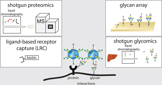
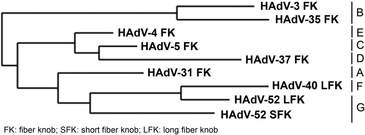
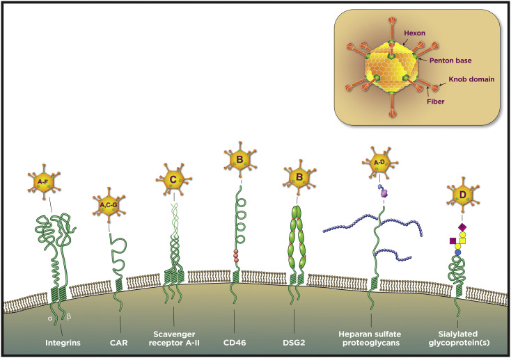
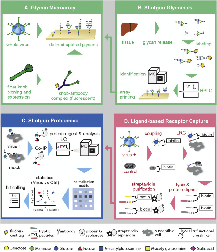
Adenoviruses as most viruses rely on glycan and protein interactions to attach to and enter susceptible host cells. The Adenoviridae family comprises more than 80 human types and they differ in their attachment factor and receptor usage, which likely contributes to the diverse tropism of the different types. In the past years, methods to systematically identify glycan and protein interactions have advanced. In particular sensitivity, speed and coverage of mass spectrometric analyses allow for high-throughput identification of glycans and peptides separated by liquid chromatography. Also, developments in glycan microarray technologies have led to targeted, high-throughput screening and identification of glycan-based receptors. The mapping of cell surface interactions of the diverse adenovirus types has implications for cell, tissue, and species tropism as well as drug development. Here we review known adenovirus interactions with glycan- and protein-based receptors, as well as glycomics and proteomics strategies to identify yet elusive virus receptors and attachment factors. We finally discuss challenges, bottlenecks, and future research directions in the field of non-enveloped virus entry into host cells.

Keywords: adenovirus; glycomis; host cell interactions; proteomics; virus entry.


Figures

Unlabelled Image
Graphical abstract
Fig. 1
Fig. 1
Selected adenovirus types and their genetic grouping. Phylogenetic tree showing the relationship of selected HAdV fiber knob (FK) sequences. HAdV-31, -3 and -35, -5, -37, -4, -40, and -52 are representing HAdV species A to G, respectively. HAdV FK sequences were obtained from NCBI. The phylogenetic tree was generated by using phylogeny.fr (http://www.phylogeny.fr/index.cgi) .
Fig. 2
Fig. 2
Adenovirus structure and important adenovirus attachment factors/receptors. (A) The adenovirus particle is 90 to 100 nm in diameter and characterized by icosahedral symmetry with a triangulation number of 25. The capsid proteins critical for cell surface binding are shown. Each of the 240 hexons comprises three identical capsid proteins. Twelve penton bases are located at the vertices. From each vertex a fiber extends and ends in a knob domain. (B) Selection of human adenovirus attachment and entry factors on the cell surface. Structures should be considered as models. The capsid component interacting with the attachment factor is indicated by the connecting line. CAR, coxsackie and adenovirus receptor; DSG2, desmoglein-2; FIX/FX, coagulation factor IX and X. Figure adapted from Arnberg .
Fig. 3
Fig. 3
Methods to identify early virus–host cell interactions. (A) Classical glycan array with synthetical glycans printed on a microarray plate. The array is probed either with purified whole virus particles, virus like particles or VAPs, such as the fiber knob domain of adenoviruses, and binding determined by fluorochromes conjugated to the probe or a probe-binding antibody. (B) Shotgun glycomics. Tissue homogenates are used to extract glycans, fluorescently label them and separate them by liquid chromatography (LC) before spotting the glycans on a microarray. The nature of each glycan can be determined by mass spectrometry (MS) while virus binding experiments are performed as in panel A. (C) Shotgun proteomics of co-immunoprecipitated virus–receptor complexes. Virus is cold-bound to cells and virus attachment protein–receptor complexes purified from cell lysates by affinity enrichment. Purified proteins are then identified and quantified by peptide fingerprinting and label-free interaction proteomics. (D) Ligand-based receptor capture uses specifically designed trifunctional organic compounds such as TRICEPS. These first react with free amines of virus surface proteins such as the fiber knob of adenoviruses and then crosslink glycosylated cell surface receptors by a hydrazine reactive group. Finally, a biotin group allows for efficient capture of receptor peptides after tryptic digest of cell lysates using streptavidin (SA) resins. Captured peptides are identified and quantified by MS as in panel C.

References

    1. Grove J., Marsh M. The cell biology of receptor-mediated virus entry. J. Cell Biol. 2011;195:1071–1082. - PMC - PubMed
    1. Amara A., Mercer J. Viral apoptotic mimicry. Nat. Rev. Microbiol. 2015;13:461–469. - PMC - PubMed
    1. Vol. 2. 2015. Principles of Virology: Pathogenesis and Control. S. Jane Flint.
    1. Marsh M., Helenius A. Virus entry: open sesame. Cell. 2006;124:729–740. - PMC - PubMed
    1. Arnberg N. Adenovirus receptors: implications for targeting of viral vectors. Trends Pharmacol. Sci. 2012;33:442–448. - PubMed

Publication types

LinkOut - more resources