. 2018 Jul 15:383:33-45.
doi: 10.1016/j.neuroscience.2018.04.036. Epub 2018 May 8.

Cofilin Knockdown Attenuates Hemorrhagic Brain Injury-induced Oxidative Stress and Microglial Activation in Mice

Affiliations

Cofilin Knockdown Attenuates Hemorrhagic Brain Injury-induced Oxidative Stress and Microglial Activation in Mice

Qasim Alhadidi et al. Neuroscience. .

Abstract

Intracerebral hemorrhage (ICH) resulting from the rupture of the blood vessels in the brain is associated with significantly higher mortality and morbidity. Clinical studies focused on alleviating the primary injury, hematoma formation and expansion, were largely ineffective, suggesting that secondary injury-induced inflammation and the formation of reactive species also contribute to the overall injury process. In this study, we explored the effects of cofilin knockdown in a mouse model of ICH. Animals given stereotaxic injections of cofilin siRNA, 72-h prior to induction of ICH by collagenase injection within the area of siRNA administration showed significantly decreased cofilin expression levels and lower hemorrhage volume and edema, and the animals performed significantly better in neurobehavioral tasks i.e., rotarod, grip strength and neurologic deficit scores. Cofilin siRNA knocked-down mice had reduced ICH-induced DNA fragmentation, blood-brain barrier disruption and microglial activation, with a concomitant increase in astrocyte activation. Increased expression of pro-survival proteins and decreased markers of oxidative stress were also observed in cofilin siRNA-treated mice possibly due to the reduced levels of cofilin. Our results suggest that cofilin plays a major role in ICH-induced secondary injury, and could become a potential therapeutic target.

Keywords: cofilin; inflammation; intracerebral hemorrhage; microglial activation; oxidative stress.


Figures

Fig. 1.
Fig. 1.
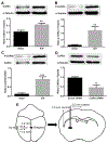
Cofilin dynamics during ICH, and cofilin siRNA transfection efficiency. ICH was induced in mice by injecting 0.09 units of collagenase in the striatum. Three days later, mice were sacrificed, brains were removed, and the tissue surrounding the hemorrhagic area was extracted and analyzed by WB. (A) Total cofilin expression was significantly increased by ICH. (B) Phospho-cofilin expression was also significantly increased by ICH. (C) The ratio of phospho-cofilin to that of total cofilin was significantly higher in the ICH group. (D) siRNA transfection mixture (siRNA mixed with Entranster-in vivo transfection reagent) was injected in the striatum. Three days later, mice were sacrificed, brains were removed and the brain tissue (1 mm around the injection site) was extracted and analyzed by WB. Cofilin siRNA significantly reduced total cofilin expression relative to the scrambled siRNA-treated group. (E) The image shows collagenase and siRNA injection sites as stated by the stereotaxic injection coordinates. N = 3–4 mice per group, unpaired t-test was used for analysis. Data are expressed as mean ± SEM, where p < 0.05 was considered significant. **p ≤ 0.01, ***p ≤ 0.001.
Fig. 2.
Fig. 2.
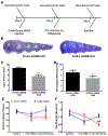
Cofilin knockdown reduced hemorrhagic volume and neurobehavioral parameters. (A) The outline shows the experimental design. Mice were trained on the rotarod and grip strength 3–7 days before siRNA injection. Baseline readings were recorded just before siRNA injection (day 0), 3 days after siRNA injection (day 3), and 3 days after ICH induction (day 6, prior to sacrifice). (B and C) Hemorrhagic injury volume analysis showed a highly significant reduction of the injury volume by cofilin siRNA relative to that of control, scrambled siRNA (n = 12 mice per group, unpaired t-test). (D) Neurological deficit scoring shows significant amelioration of neurological deficit by cofilin knockdown (n = 12 mice per group, unpaired t-test). (E) The reduction in the rotarod latency time 3 days following ICH induction was significantly improved by cofilin knockdown (n = 12 mice per group, repeated measure ANOVA followed by Bonferroni’s post hoc comparisons). (F) The reduction in grip strength 3 days following ICH induction was significantly improved by cofilin knockdown (n = 12 mice per group, repeated measure ANOVA followed by Bonferroni’s post hoc comparisons). Data are expressed as mean ± SEM, where the p < 0.05 was considered significant. *Difference between groups. #Difference within groups relative to baseline. *p ≤ 0.05, **p ≤ 0.01, #p ≤ 0.05, ###p ≤ 0.001.
Fig. 3.
Fig. 3.
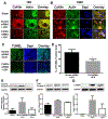
Cofilin siRNA reduced cofilin expression and apoptosis after ICH. (A) Immunohistochemical staining of ICH mice brain sections showed that cofilin expression was highly increased within and around the hemorrhagic area, and cofilin knockdown reduced this effect (n = 3–4 mice per group). (B) Cofilin–actin aggregates illustrated at 100× magnification were abundant in the cells around the hemorrhagic area. Cofilin knockdown reduced aggregate formation in these cells (n = 3–4 mice per group). (C) Cofilin knockdown reduced the number of TUNEL-positive cells induced by ICH. (D) Data analysis for the percent of TUNEL-positive cells showed significant reduction in TUNEL-positive cells by cofilin knockdown (n = 4 mice per group, unpaired t-test). (E) WB analysis of active cleaved caspase 3 showed that cofilin knockdown reduced activation of caspase 3 induced by ICH (n = 3–4 mice per group, one-way ANOVA). (F) WB analysis of total caspase 3 showed no reduction in the expression of the total caspase 3 among sham, scram siRNA and cofilin siRNA groups (n = 3–4 mice per group, one-way ANOVA). (G) WB analysis of cleaved PARP showed reduced trend in cofilin siRNA-treated group (n = 3–4 mice per group, one-way ANOVA). Data are expressed as mean ± SEM, where the p ≤ 0.05 was considered significant. *p ≤ 0.05.
Fig. 4.
Fig. 4.
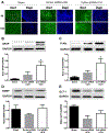
Cofilin knockdown reduced glial cell activation after ICH. (A) ICH induced the activation of microglial cells around the hemorrhagic area illustrated by immunohistochemical staining of Iba1, a marker of microglial cell activation. Cofilin knockdown reduced Iba1 expression and consequently microglial cell activation (n = 3–4 mice per group). (B) ICH slightly induced the activation of astrocytes illustrated by WB analysis of GFAP, a marker of astrocyte activation but was significantly increased by cofilin knockdown (n = 3–4 mice per group, one-way ANOVA). (C) Akt activation was significantly increased by cofilin knockdown (n = 3–4 mice per group, one-way ANOVA). (D) BDNF expression showed an increased trend in its levels in the cofilin siRNA-treated group (n = 3–4 mice per group, one-way ANOVA). (E) GLT-1 expression also showed an increased trend in the cofilin siRNA-treated group (n = 3–4 mice per group, one-way ANOVA). Data are expressed as mean ± SEM, where p ≤ 0.05 was considered significant. *p ≤ 0.05.
Fig. 5.
Fig. 5.
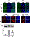
Cofilin knockdown reduced oxidative/nitrosative stress after ICH. (A and B) The oxidative stress manifestation illustrated by immunohistochemical staining of the oxidized nucleoside 8-HG was highly increased after ICH. Cofilin knockdown reduced the level of 8-HG at and around the hemorrhagic area visualized at 10× magnification (A) and 100× magnification (B). The co-localization of 8-HG with DAPI was clear at higher magnifications only (n = 3–4 mice per group). (C and D) Protein nitration evaluated by immunohistochemical staining as well as WB analysis of 3-NT was also increased after ICH. Cofilin knockdown reduced the levels of nitrated proteins within the hemorrhagic area visualized at 4× and 10× magnifications (C) (n = 3–4 mice per group). Nitrated protein levels were significantly reduced by cofilin knockdown (D), (n = 4 mice per group). Data are expressed as mean ± SEM and p ≤ 0.05 was considered significant. *p ≤ 0.05.
Fig. 6.
Fig. 6.
Cofilin knockdown reduced ICH-induced edema and BBB disruption. (A) Water content in the ipsilateral striatum was significantly reduced by cofilin knockdown. No significant differences were detected in other regions (n = 7 mice per group). (B and C) BBB disruption was evaluated by fluorescence imaging of the extravasated FITC-labeled dextran particles in the hemorrhagic area. Starting from 1 mm before the hemorrhagic area, 4 sections taken from different regions were selected and imaged using fluorescent microscopy (B). The average fluorescence intensity for each section was quantified using image J software and expressed as integrated density. Although the reduction in integrated density triggered by cofilin siRNA treatment was statistically not significant (C), however, there was a trend supporting the water content data (n = 3 mice per group, unpaired t-test). Data are expressed as mean ± SEM and p ≤ 0.05 was considered significant. *p ≤ 0.05.
Fig. 7.
Fig. 7.
The proposed mechanism of cofilin signaling during ICH. Thrombin released during hemorrhage is the major activator of Rho GTPases in different cell types. Subsequently, cofilin activation leads to neuronal apoptosis, glial activation, and BBB disruption of microvascular endothelial cells.

References

    1. Adeoye O, Broderick JP (2010) Advances in the management of intracerebral hemorrhage. Nat Rev Neurol 6:593–601. - PubMed
    1. Alhadidi Q, Bin Sayeed MS, Shah ZA (2016) Cofilin as a promising therapeutic target for ischemic and hemorrhagic stroke. Transl Stroke Res 7:33–41. - PubMed
    1. Alhadidi Q, Sayeed MS, Shah Z (2017) The interplay between cofilin and phospho-cofilin: its role in maintaining blood brain barrier integrity. CNS Neurol Disord Drug Targets. - PubMed
    1. Alhadidi Q, Shah ZA (2017) Cofilin mediates LPS-induced microglial cell activation and associated neurotoxicity through activation of NF-kappaB and JAK-STAT pathway. Mol Neurobiol. - PMC - PubMed
    1. Aronowski J, Zhao X (2011) Molecular pathophysiology of cerebral hemorrhage: secondary brain injury. Stroke 42:1781–1786. - PMC - PubMed

Publication types

Substances

LinkOut - more resources