. 2018 Jun 1;17(6):2216-2225.
doi: 10.1021/acs.jproteome.8b00180. Epub 2018 May 18.

Multiproteomic and Transcriptomic Analysis of Oncogenic β-Catenin Molecular Networks

Affiliations

Multiproteomic and Transcriptomic Analysis of Oncogenic β-Catenin Molecular Networks

Rob M Ewing et al. J Proteome Res. .

Abstract

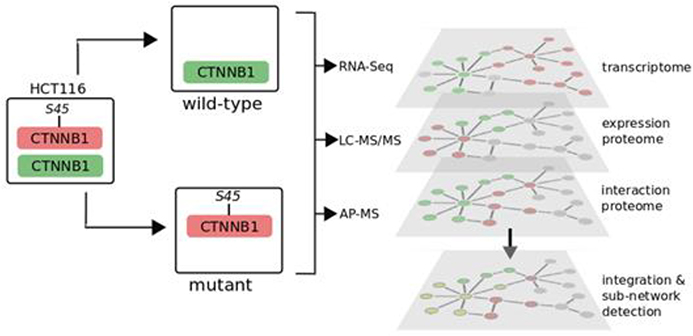
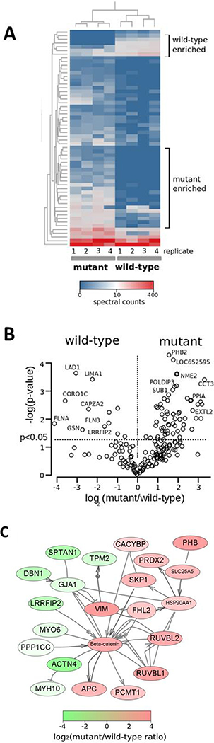
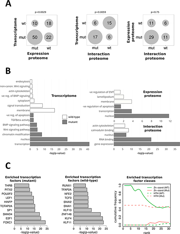
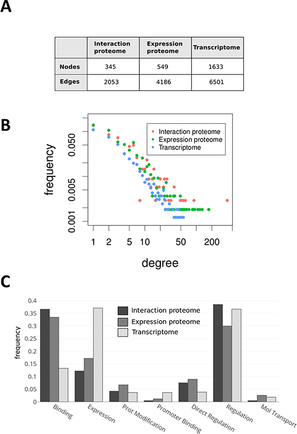
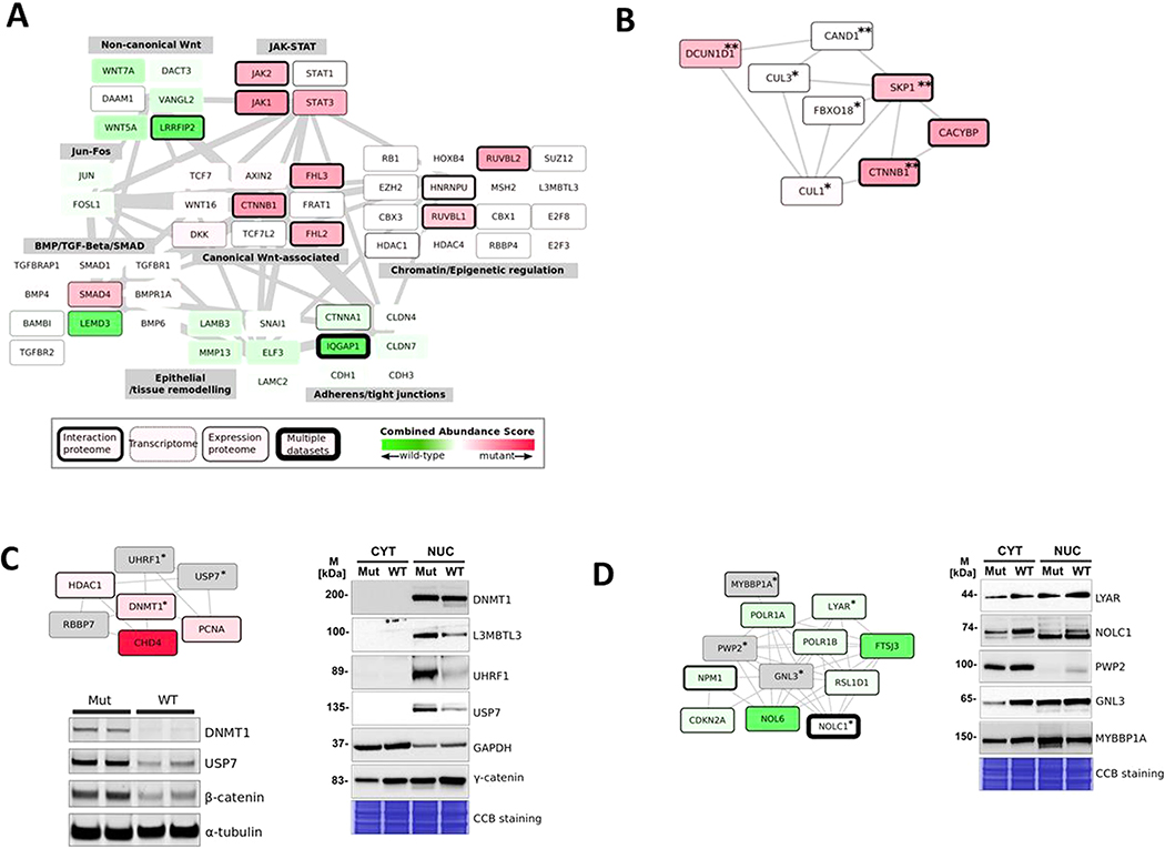
The dysregulation of Wnt signaling is a frequent occurrence in many different cancers. Oncogenic mutations of CTNNB1/β-catenin, the key nuclear effector of canonical Wnt signaling, lead to the accumulation and stabilization of β-catenin protein with diverse effects in cancer cells. Although the transcriptional response to Wnt/β-catenin signaling activation has been widely studied, an integrated understanding of the effects of oncogenic β-catenin on molecular networks is lacking. We used affinity-purification mass spectrometry (AP-MS), label-free liquid chromatography-tandem mass spectrometry, and RNA-Seq to compare protein-protein interactions, protein expression, and gene expression in colorectal cancer cells expressing mutant (oncogenic) or wild-type β-catenin. We generate an integrated molecular network and use it to identify novel protein modules that are associated with mutant or wild-type β-catenin. We identify a DNA methyltransferase I associated subnetwork that is enriched in cells with mutant β-catenin and a subnetwork enriched in wild-type cells associated with the CDKN2A tumor suppressor, linking these processes to the transformation of colorectal cancer cells through oncogenic β-catenin signaling. In summary, multiomics analysis of a defined colorectal cancer cell model provides a significantly more comprehensive identification of functional molecular networks associated with oncogenic β-catenin signaling.

Keywords: DNA methyltransferase I; Wnt signaling; multiomics integration; oncogenic mutations; protein−protein interaction network; β-catenin.


Conflict of interest statement

The authors declare no competing financial interest.

Figures

Figure 1.
Figure 1.
Integrated multiomics analysis of β-catenin signaling networks. Experimental design and data acquisition of interactome (AP-MS), expression proteome (LC–MS/MS), and transcriptome (RNA-Seq) from colorectal cancer cell lines HCT116-CTNNB1−/Δ45 (mutant) and HCT116-CTNNB1WT/− (wild-type) expressing endogenous mutant or wild-type CTNNB1/β-catenin
Figure 2.
Figure 2.
AP-MS analysis of mutant and wild-type β-catenin protein interactions. (A) Heatmap of protein spectral counts across four replicate HCT116-CTNNB1−/Δ45 (mutant) and four HCT116-CTNNB1WT/− (wild-type) AP-MS samples. Selected profiles of proteins associated with either mutant AP-MS or wild-type AP-MS samples are shown. (B) Volcano plot indicating log2 ratio of mutant-to-wild-type spectral counts from a single AP-MS study, with significant (p < 0.05) proteins indicated. (C) Network diagram of β-catenin (CTNNB1) interaction partners identified in the study. The largest connected component subnetwork in the Pathway Studio analysis is shown. Proteins are shaded according to their mutant-to-wild-type spectral count ratio (red-shaded proteins are highly enriched in mutant AP-MS samples, and green-shaded proteins are highly enriched in wild-type AP-MS samples).
Figure 3.
Figure 3.
Functional analysis of β-catenin-associated proteomic and transcriptomic profiles (A) Bubble plot indicating the size of the intersections of Gene Ontology (GO) terms between interaction, expression-proteome, and transcriptomic data sets. The numbers indicate shared GO terms for each comparison for GO terms significantly (p < 0.05) enriched in mutant or wild-type samples. The p values are Fisher’s exact test values indicating the significance of the observed overlap of GO terms.(B) Enriched GO terms in mutant and wild-type cells across each data set. The most significantly differential GO terms were identified for each data set by comparing the p values for each term between mutant and wild-type gene sets. (C) Enriched transcription factors in the significantly (p < 0.05) differential mutant or wild-type gene sets from RNA-Seq analysis. Enrichr analysis was used to identify the most-enriched transcription factors in the significantly different (p < 0.05) RNA-Seq data sets. The top 10 enriched transcription factors are shown for mutant and wild-type samples (panel 1 and 2). Ranked transcription factor classes for the mutant and wild-type RNA-Seq significantly different data sets show distinct classes of transcription factors in each cell type.
Figure 4.
Figure 4.
Network properties of proteomic and transcriptomic data sets. (A) Summary of network properties from the integrated network constructed by integrating all three data sets with known protein–protein interactions. The table indicates the numbers of nodes (protein and genes) and edges (relations between proteins) from each data set integrated into the combined network. (B) Log–log plot of the degree distributions for nodes from each data set (the number of connections for protein nodes typically follow the pattern of interaction proteome > expression proteome > transcriptome). (C) Analysis of interaction (edge) types for each data set indicate significant differences of functional type of edges contributed to the integrated network.
Figure 5.
Figure 5.
Integrated proteomic and transcriptomic functional modules. (A) Selected functional modules from the integrated network. Edge thickness represents the overall connectivity between modules (normalized edge weights calculated as the total number of edges divided by the number of genes and proteins in each module). Node (gene and protein) color intensity indicates the combined abundance score (red, mutant; green, wild-type). (B) SCF (Skp-Cullin-F-box)-associated protein network showing proteins significantly (double asterisks indicate p < 0.05; a single asterisk indicates p < 0.1) abundant in interaction- and expression-proteome data sets. (C) DNA methyltransferase I (Dnmt1)-associated protein network. Protein nodes marked with an asterisk were also tested by immunoblotting as shown. Dnmt1, USP7, and β-catenin were tested using immunoblotting on whole-cell lysates from mutant and wild-type cells and additional related interaction partners (UHRF1, L3MBTL3) analyzed by the immunoblotting of nuclear and cytosolic subcellular fractions from mutant and wild-type cells. (D) Western analysis of the ribosome-biogenesis-associated protein network in subcellular fractionated samples. Protein nodes marked with asterisks were also tested by immunoblotting, as shown in nuclear and cytosolic fractions from mutant and wild-type cells as in Figure 5C.

References

    1. Clevers H Wnt/Beta-Catenin Signaling in Development and Disease. Cell 2006, 127 (3), 469–480. - PubMed
    1. Amit S; Hatzubai A; Birman Y; Andersen JS; Ben-Shushan E; Mann M; Ben-Neriah Y; Alkalay I Axin-Mediated CKI Phosphorylation of Beta-Catenin at Ser 45: A Molecular Switch for the Wnt Pathway. Genes Dev. 2002, 16 (9), 1066–1076. - PMC - PubMed
    1. Morin PJ; Sparks AB; Korinek V; Barker N; Clevers H; Vogelstein B; Kinzler KW Activation of Beta-Catenin-Tcf Signaling in Colon Cancer by Mutations in Beta-Catenin or APC. Science 1997, 275 (5307), 1787–1790. - PubMed
    1. Valenta T; Hausmann G; Basler K The Many Faces and Functions of β-Catenin. EMBO J. 2012, 31 (12), 2714–36. - PMC - PubMed
    1. Angers S; Moon RT Proximal Events in Wnt Signal Transduction. Nat. Rev. Mol. Cell Biol. 2009, 10, 468. - PubMed

Publication types

MeSH terms

Substances