. 2018 May 10;19(1):351.
doi: 10.1186/s12864-018-4738-2.

Functional signaling and gene regulatory networks between the oocyte and the surrounding cumulus cells

Affiliations

Functional signaling and gene regulatory networks between the oocyte and the surrounding cumulus cells

Fernando H Biase et al. BMC Genomics. .

Abstract

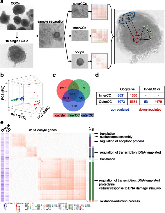
Background: The maturation and successful acquisition of developmental competence by an oocyte, the female gamete, during folliculogenesis is highly dependent on molecular interactions with somatic cells. Most of the cellular interactions identified, thus far, are modulated by growth factors, ions or metabolites. We hypothesized that this interaction is also modulated at the transcriptional level, which leads to the formation of gene regulatory networks between the oocyte and cumulus cells. We tested this hypothesis by analyzing transcriptome data from single oocytes and the surrounding cumulus cells collected from antral follicles employing an analytical framework to determine interdependencies at the transcript level.

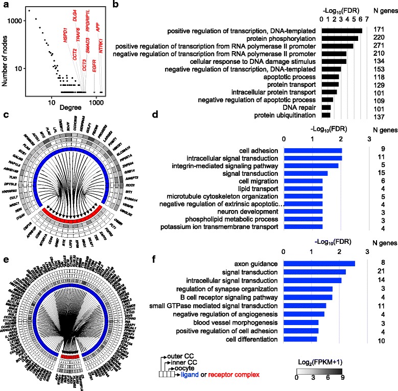
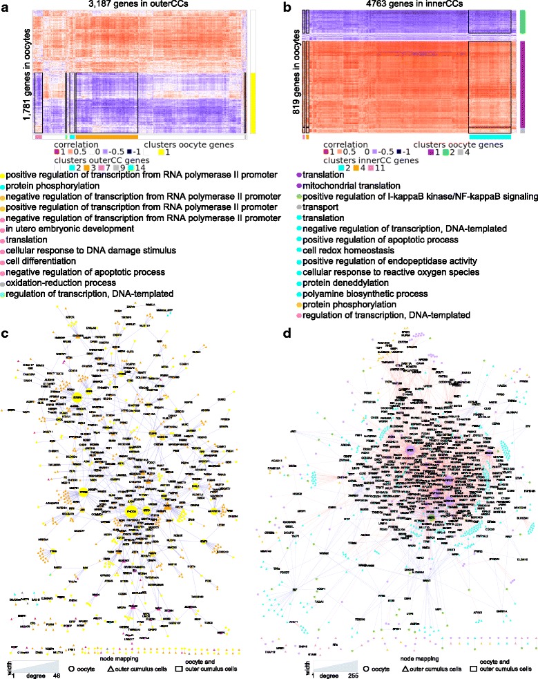
Results: We overlapped our transcriptome data with putative protein-protein interactions and identified hundreds of ligand-receptor pairs that can transduce paracrine signaling between an oocyte and cumulus cells. We determined that 499 ligand-encoding genes expressed in oocytes and cumulus cells are functionally associated with transcription regulation (FDR < 0.05). Ligand-encoding genes with specific expression in oocytes or cumulus cells were enriched for biological functions that are likely associated with the coordinated formation of transzonal projections from cumulus cells that reach the oocyte's membrane. Thousands of gene pairs exhibit significant linear co-expression (absolute correlation > 0.85, FDR < 1.8 × 10- 5) patterns between oocytes and cumulus cells. Hundreds of co-expressing genes showed clustering patterns associated with biological functions (FDR < 0.5) necessary for a coordinated function between the oocyte and cumulus cells during folliculogenesis (i.e. regulation of transcription, translation, apoptosis, cell differentiation and transport).

Conclusion: Our analyses revealed a complex and functional gene regulatory circuit between the oocyte and surrounding cumulus cells. The regulatory profile of each cumulus-oocyte complex is likely associated with the oocytes' developmental potential to derive an embryo.

Keywords: Gametogenesis; Gene regulatory networks; Inter-cellular communication; Oocyte-cumulus signaling.


Conflict of interest statement

Ethics approval and consent to participate

Ovaries were purchased from Brown Packing, SC. There was no handling of live animals for this experiment and ovaries were obtained postmortem. The Public Health Service Policy on Humane Care and Use of Laboratory Animals does not cover the use of parts of dead animals, thus no IACUC approval was needed for this experiment.

Competing interests

The authors declare they have not competing interests.

Publisher’s Note

Springer Nature remains neutral with regard to jurisdictional claims in published maps and institutional affiliations.

Figures

Fig. 1
Fig. 1
Transcriptome heterogeneity between oocytes and surrounding cumulus cells. (a) Depiction of the experimental design and the sequenced samples. (b) Principal component analysis of 12,482 genes quantified in oocytes (red), innerCCs (green) and outerCCs (blue). (c) Overlap of genes expressed in oocytes (red), innerCCs (green) or outerCCs (blue). (d) Differentially expressed genes determined from the pairwise comparisons of the three sample types (FDR < 0.01). (e) Co-expression analysis of genes expressed in oocytes showing differential expression compared those in cumulus cells (CC). The blue heatmaps depict the average expression in oocytes and CCs; the center heatmap depicts the topological overlap of correlated expression; and the vertical bars show the annotation of GO biological processes (BP) and molecular functions (MF) enriched in the corresponding gene cluster. The squares below the heatmap provide a link between cluster number, the corresponding biological process or molecular functions and genes presented in the supplementary (Additional file 3: Tables S8-S9)
Fig. 2
Fig. 2
Ligand-receptor pairs in oocytes and surrounding cumulus cells. (a) Distribution of ligands or receptors among genes expressed in COCs according to the degree of connectivity. (b) Biological processes enriched (FDR < 0.05) in ligands or receptors in coding genes expressed in COCs (see Additional file 3: Table S13 for full list of terms). (c) Ligand-encoding genes expressed in CCs with receptor-encoding genes expressed in oocytes. (d) Biological processes enriched (FDR < 0.05) in ligand-encoding genes expressed in CCs with receptor-encoding genes expressed in oocytes. (e) Ligand-encoding genes expressed in oocytes with receptor-encoding genes expressed in CCs. (f) Biological processes enriched (FDR < 0.05) in ligand-encoding genes expressed in oocytes with receptor-encoding genes expressed in CCs. To improve readability, genes with average FPKM > 2 were plotted on panels c and e
Fig. 3
Fig. 3
Variability in gene expression in COCs associated with biological functions. Coefficients of variation for genes expressed in all samples of oocytes (a) and outerCCs (b). Genes associated with specific categories are indicated with colored dots (P-value). The green line indicates the coefficient of variation threshold used to identify highly variable genes
Fig. 4
Fig. 4
Functional co-expression between oocytes and surrounding cumulus cells. Heatmap of genes showing a high correlation (|bicor| > 0.85) of expression values between oocytes and outerCCs (a) and oocytes and innerCCs (b). Horizontal and vertical bars next to the heatmaps annotate gene clusters with enriched biological processes (FDR < 0.2). The black boxes indicate the co-expression blocks with intersection of clusters enriched for biological processes. Co-expression networks between oocytes and outerCCs (c) and oocytes and innerCCs (d). The color of symbols in panels c and d correspond to the colored circles and the GO biological processes on panels a and b, respectively

References

    1. Diaz FJ, Wigglesworth K, Eppig JJ. Oocytes are required for the preantral granulosa cell to cumulus cell transition in mice. Dev Biol. 2007;305(1):300–311. doi: 10.1016/j.ydbio.2007.02.019. - DOI - PMC - PubMed
    1. Conti M, Hsieh M, Zamah AM, Oh JS. Novel signaling mechanisms in the ovary during oocyte maturation and ovulation. Mol Cell Endocrinol. 2012;356(1–2):65–73. doi: 10.1016/j.mce.2011.11.002. - DOI - PMC - PubMed
    1. Amireault P, Dube F. Intracellular cAMP and calcium signaling by serotonin in mouse cumulus-oocyte complexes. Mol Pharmacol. 2005;68(6):1678–1687. - PubMed
    1. Gilchrist RB, Ritter LJ, Armstrong DT. Oocyte-somatic cell interactions during follicle development in mammals. Anim Reprod Sci. 2004;82-3:431–446. doi: 10.1016/j.anireprosci.2004.05.017. - DOI - PubMed
    1. Wigglesworth K, Lee KB, O'Brien MJ, Peng J, Matzuk MM, Eppig JJ. Bidirectional communication between oocytes and ovarian follicular somatic cells is required for meiotic arrest of mammalian oocytes. P Natl Acad Sci USA. 2013;110(39):E3723–E3729. doi: 10.1073/pnas.1314829110. - DOI - PMC - PubMed