. 2018 May 10;6(1):34.
doi: 10.1186/s40425-018-0347-5.

CAR T cell therapy for breast cancer: harnessing the tumor milieu to drive T cell activation

Affiliations

CAR T cell therapy for breast cancer: harnessing the tumor milieu to drive T cell activation

Pradip Bajgain et al. J Immunother Cancer. .

Abstract

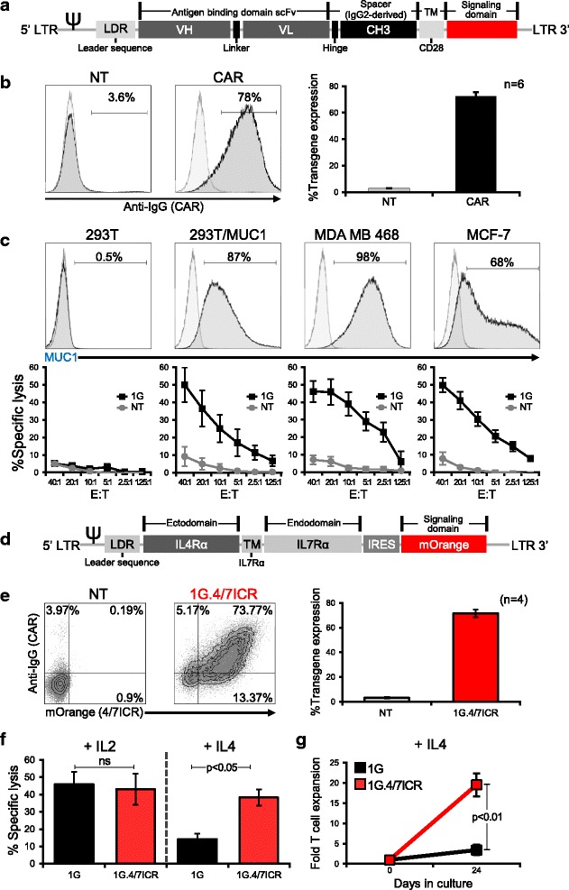
Background: The adoptive transfer of T cells redirected to tumor via chimeric antigen receptors (CARs) has produced clinical benefits for the treatment of hematologic diseases. To extend this approach to breast cancer, we generated CAR T cells directed against mucin1 (MUC1), an aberrantly glycosylated neoantigen that is overexpressed by malignant cells and whose expression has been correlated with poor prognosis. Furthermore, to protect our tumor-targeted cells from the elevated levels of immune-inhibitory cytokines present in the tumor milieu, we co-expressed an inverted cytokine receptor linking the IL4 receptor exodomain with the IL7 receptor endodomain (4/7ICR) in order to transform the suppressive IL4 signal into one that would enhance the anti-tumor effects of our CAR T cells at the tumor site.

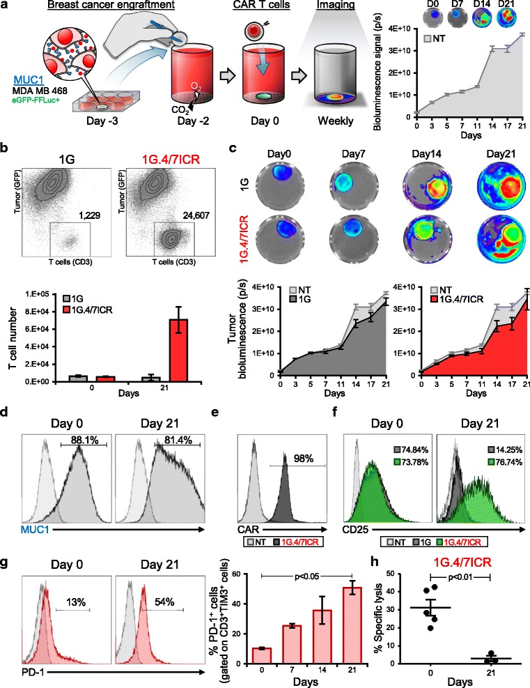
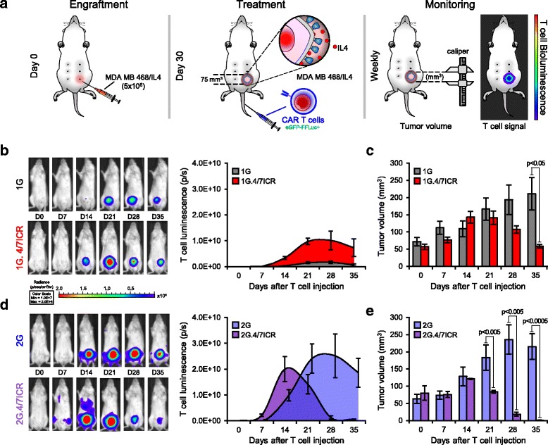
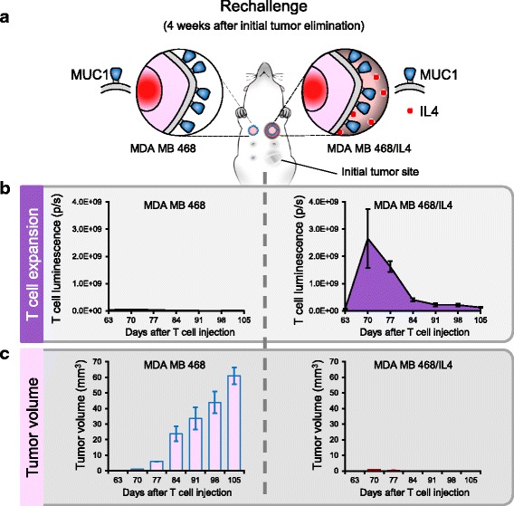
Methods: First (1G - CD3ζ) and second generation (2G - 41BB.CD3ζ) MUC1-specific CARs were constructed using the HMFG2 scFv. Following retroviral transduction transgenic expression of the CAR±ICR was assessed by flow cytometry. In vitro CAR/ICR T cell function was measured by assessing cell proliferation and short- and long-term cytotoxic activity using MUC1+ MDA MB 468 cells as targets. In vivo anti-tumor activity was assessed using IL4-producing MDA MB 468 tumor-bearing mice using calipers to assess tumor volume and bioluminescence imaging to track T cells.

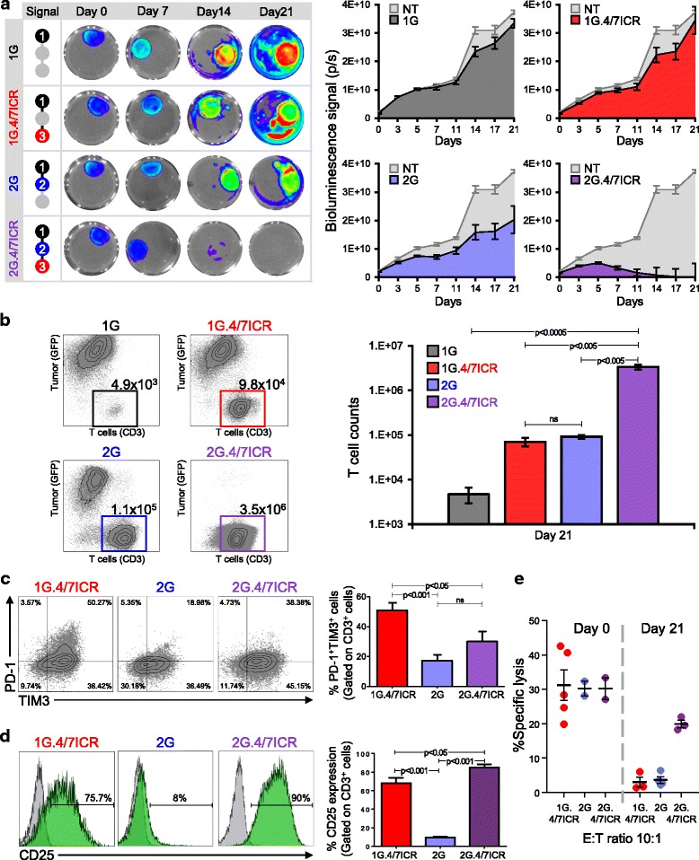
Results: In the IL4-rich tumor milieu, 1G CAR.MUC1 T cells failed to expand or kill MUC1+ tumors and while co-expression of the 4/7ICR promoted T cell expansion, in the absence of co-stimulatory signals the outgrowing cells exhibited an exhausted phenotype characterized by PD-1 and TIM3 upregulation and failed to control tumor growth. However, by co-expressing 2G CAR.MUC1 (signal 1 - activation + signal 2 - co-stimulation) and 4/7ICR (signal 3 - cytokine), transgenic T cells selectively expanded at the tumor site and produced potent and durable tumor control in vitro and in vivo.

Conclusions: Our findings demonstrate the feasibility of targeting breast cancer using transgenic T cells equipped to thrive in the suppressive tumor milieu and highlight the importance of providing transgenic T cells with signals that recapitulate physiologic TCR signaling - [activation (signal 1), co-stimulation (signal 2) and cytokine support (signal 3)] - to promote in vivo persistence and memory formation.

Keywords: Breast cancer; Chimeric antigen receptor; Genetic engineering; Inverted cytokine receptor; T cell therapy.


Conflict of interest statement

Ethics approval and consent to participate

Collection of human peripheral blood mononuclear cells (PBMCs) were obtained from healthy donors and patients after informed consent on protocols approved by the Institutional Review Board (IRB) at Baylor College of Medicine (H-15152). Mice were housed and treated in accordance with Baylor College of Medicine Animal Husbandry and Institutional Animal Care and Use Committee guidelines (AN-5639).

Competing interests

The Center for Cell and Gene Therapy at Baylor College of Medicine established a collaboration with Celgene at the time the work was conducted. S.S., A.M.L., and J.F.V. have filed a related patent application.

Publisher’s Note

Springer Nature remains neutral with regard to jurisdictional claims in published maps and institutional affiliations.

Figures

Fig. 1
Fig. 1
4/7ICR improves the cytolytic function and proliferation of CAR.MUC1 T cells in presence of IL4. a Schematic representation of 1st generation CAR.MUC1 (1G) construct. b CAR.MUC1 expression on activated T cells measured 3 days post-transduction (representative donor on the left, summary data on the right). Data represents mean ± SEM (n = 6). c Phenotypic analysis of MUC1 expression on different cell lines (top panel) and in vitro cytolytic function of control (NT) and CAR T cells assessed in a 5 hr 51Cr-release assay at E:Ts of 1.25:1 to 40:1, using MUC1+ targets (293T/MUC1, MDA MB 468, MCF-7) and MUC1- target (293T) (bottom). Data represents mean ± SEM (n = 5). d Schematic of 4/7ICR vector map. e Transgenic expression of both 4/7ICR and CAR.MUC1 in T cells as detected by mOrange and anti-IgG, respectively. Right panel shows summary data representing the percentage of double-positive cells (1G.4/7ICR) (mean ± SEM, n = 4). f Cytolytic function of transgenic (1G or 1G.4/7ICR) T cells pre-exposed to IL4 as assessed in a 4 hr 51Cr-release assay using MDA MB 468 as a target at the indicated E:T ratios. Statistical significance was calculated between 1G and 1G.4/7ICR using One-way ANOVA, p < 0.05. g Cell expansion of 1G or 1G.4/7ICR T cells (1 × 106) stimulated weekly with irradiated MDA MB 468 cells (0.5 × 106) with IL4 (400 U/mL) added twice weekly. T cell expansion was quantified by cell counting using trypan blue exclusion to assess cell viability. Statistical significance was calculated between 1G and 1G.4/7ICR using One-way ANOVA, p < 0.01
Fig. 2
Fig. 2
Transgenic expression of 4/7ICR is insufficient to overcome tumor-mediated T cell dysfunction. a Schematic of co-culture experimental setup (left panel) and bioluminescence data tracking tumor expansion (in the absence of T cell treatment) over time (right panel). b Representative dot plots (top) and summary quantitative data (bottom) showing T cells and tumor cell numbers on day 0 and day 21 of coculture (mean ± SEM, n = 6 independent experiments). Significance was determined by an unpaired two-tailed t-test, p < 0.05, 1G.4/7ICR compared with 1G. c Representative images of bioluminescent tumor cells (top) and summarized quantitative bioluminescence signal from tumor cells treated with either NT, 1G or 1G.4/7ICR T cells over time (mean ± SEM, n = 6). d Phenotypic analysis of MUC1 expression on MDA MB 468 cells on day 0 and day 21 of co-culture (representative donor). e CAR expression on 1G.4/7ICR cells after co-culture with tumor cells. f CD25 expression on 1G versus 1G.4/7ICR T cells on day 0 and day 21 of co-culture (light grey: isotype control, dark grey: 1G T cells, green: 1G.4/7ICR T cells). g Surface expression of PD-1 (representative donor - left, summary data - right) on 1G.4/7ICR cells gated on CD3+TIM3+cells and analyzed on days 0, 7, 14 and 21 of co-culture (mean ± SEM, n = 6, p < 0.05). h Cytolytic activity of 1G.4/7/ICR prior to and day 21 post-coculture using MDA MB 468 cells as targets (E:T 10:1; mean ± SEM, n = 4, p < 0.01)
Fig. 3
Fig. 3
Combining 4/7ICR with a 2G CAR preserves T cell function even under suppressive conditions. a Serial bioluminescence imaging of eGFP-FFLuc+ MDA MB 468 cells co-cultured with 1G, 1G.4/7ICR, 2G, or 2G.4/7ICR T cells in the presence of IL4 [representative images - left, quantitative data - right (n = 6)]. b Representative dot plots and summary FACS data quantifying tumor cells and T cells after 21 days of co-culture. c Surface expression of PD-1 and TIM3 on transgenic T cells analyzed on day 21 after co-culture (representative data - left, summary data - right). d CD25 expression on 1G.4/7ICR, 2G, and 2G.4/7ICR cells on day 21 of coculture. e In vitro cytolytic function of 1G.4/7ICR, 2G, and 2G.4/7ICR cells isolated on day 21 after co-culture (mean ± SEM, n = 4–6). Significance was determined by two-way ANOVA. p < 0.05, p < 0.01, p < 0.001
Fig. 4
Fig. 4
Combined expression of 4/7ICR and 2G CAR augments anti-tumor activity in vivo. a Schematic of in vivo experiment where NSG mice with IL4-producing MDA MB 468 cells and treated i.v. with eGFP-FFLuc+1G, 1G.4/7ICR, 2G, or 2G.4/7ICR T cells. b Representative animal images (left) and summary bioluminescence data (right, mean ± SEM, n = 3–5/group) indicating T cell localization and expansion. c Tumor volume measured by calipers (mean ± SEM, n = 3–5/group). Significance was determined by two-way ANOVA. p < 0.05 on day 35. d Representative animal images (left) and summary bioluminescence data (right, mean ± SEM, n = 3–5/group). e Tumor volume measured by calipers. Significance was determined by two-way ANOVA
Fig. 5
Fig. 5
2G.4/7ICR T cells persist long term and retain their anti-tumor activity and tumor selectivity. a Schema of tumor rechallenge model. b T cell expansion over time as assessed by quantitative bioluminescence imaging at the site of MDA MB 468 (left) and MDA MB 468/IL4 (right) tumor cell injection. c Tumor volume as measured by calipers

References

    1. Siegel RL, Miller KD, Jemal A. Cancer statistics, 2017. CA Cancer J Clin. 2017;67(1):7–30. doi: 10.3322/caac.21387. - DOI - PubMed
    1. Brentjens RJ, Davila ML, Riviere I, Park J, Wang X, Cowell LG, Bartido S, Stefanski J, Taylor C, Olszewska M, et al. CD19-targeted T cells rapidly induce molecular remissions in adults with chemotherapy-refractory acute lymphoblastic leukemia. Sci Transl Med. 2013;5(177):177ra38. doi: 10.1126/scitranslmed.3005930. - DOI - PMC - PubMed
    1. Grupp SA, Kalos M, Barrett D, Aplenc R, Porter DL, Rheingold SR, Teachey DT, Chew A, Hauck B, Wright JF, et al. Chimeric antigen receptor-modified T cells for acute lymphoid leukemia. N Engl J Med. 2013;368(16):1509–1518. doi: 10.1056/NEJMoa1215134. - DOI - PMC - PubMed
    1. Kalos M, Levine BL, Porter DL, Katz S, Grupp SA, Bagg A, June CH. T cells with chimeric antigen receptors have potent antitumor effects and can establish memory in patients with advanced leukemia. Sci Transl Med. 2011;3(95):95ra73. doi: 10.1126/scitranslmed.3002842. - DOI - PMC - PubMed
    1. Porter DL, Levine BL, Kalos M, Bagg A, June CH. Chimeric antigen receptor-modified T cells in chronic lymphoid leukemia. N Engl J Med. 2011;365(8):725–733. doi: 10.1056/NEJMoa1103849. - DOI - PMC - PubMed

Publication types

MeSH terms

Substances