. 2018 May 31;475(10):1821-1837.
doi: 10.1042/BCJ20180171.

An allostatic mechanism for M2 pyruvate kinase as an amino-acid sensor

Affiliations

An allostatic mechanism for M2 pyruvate kinase as an amino-acid sensor

Meng Yuan et al. Biochem J. .

Abstract

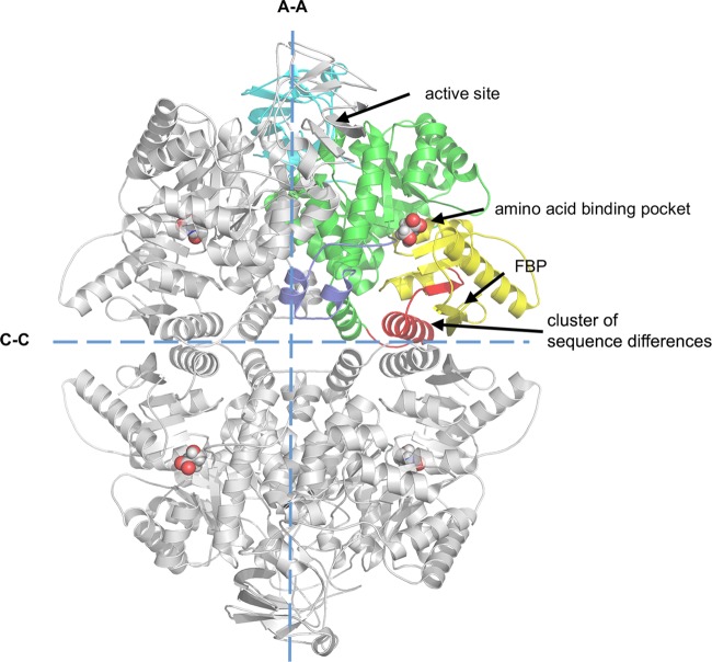
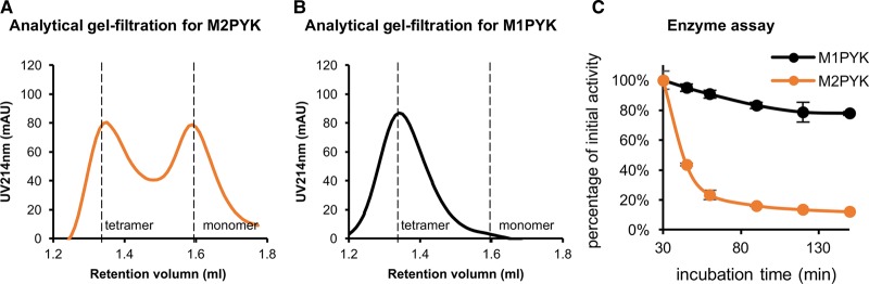
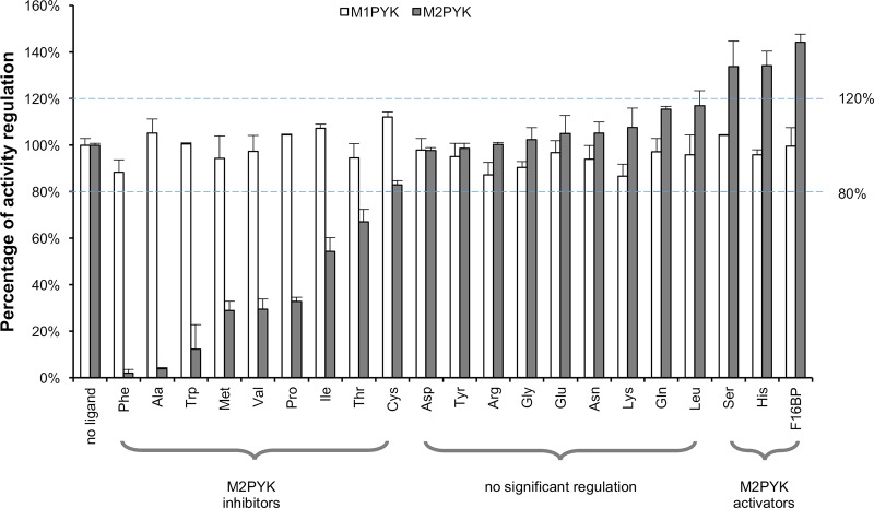
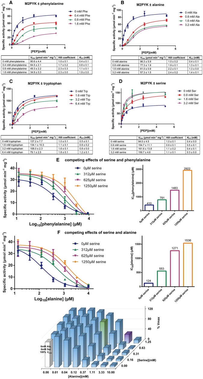
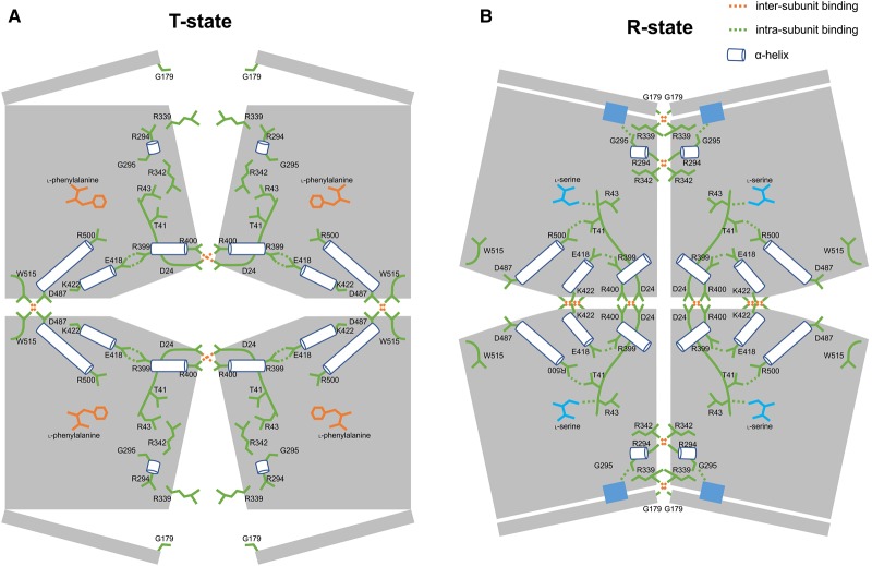
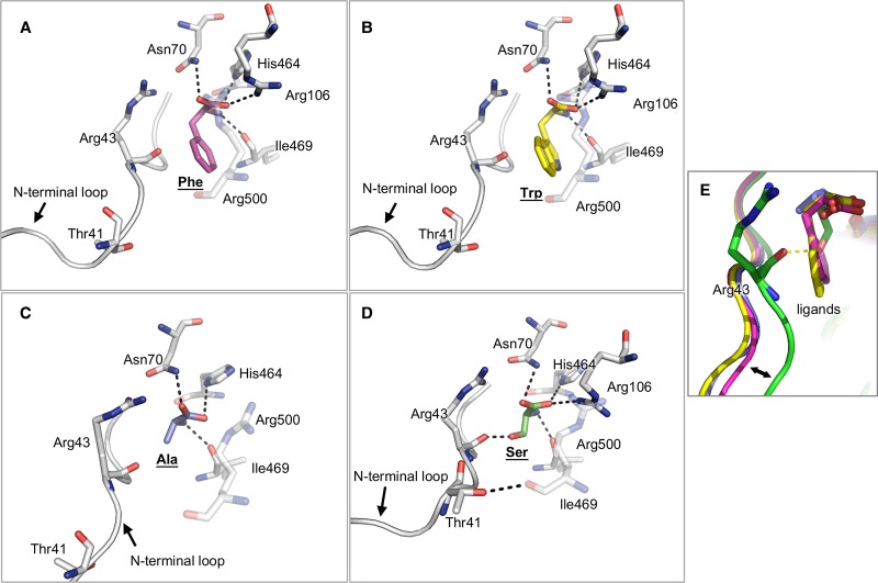
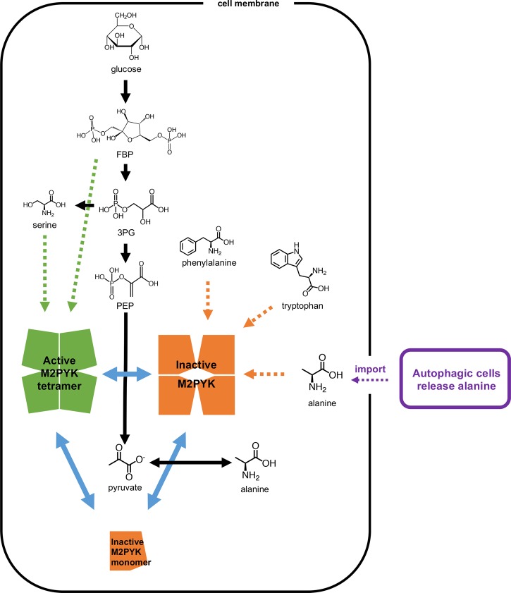
We have tested the effect of all 20 proteinogenic amino acids on the activity of the M2 isoenzyme of pyruvate kinase (M2PYK) and show that, within physiologically relevant concentrations, phenylalanine, alanine, tryptophan, methionine, valine, and proline act as inhibitors, while histidine and serine act as activators. Size exclusion chromatography has been used to show that all amino acids, whether activators or inhibitors, stabilise the tetrameric form of M2PYK. In the absence of amino-acid ligands an apparent tetramer-monomer dissociation Kd is estimated to be ∼0.9 µM with a slow dissociation rate (t1/2 15 min). X-ray structures of M2PYK complexes with alanine, phenylalanine, and tryptophan show the M2PYK locked in an inactive T-state conformation, while activators lock the M2PYK tetramer in the active R-state conformation. Amino-acid binding in the allosteric pocket triggers rigid body rotations (11°) stabilising either T or R states. The opposing inhibitory and activating effects of the non-essential amino acids serine and alanine suggest that M2PYK could act as a rapid-response nutrient sensor to rebalance cellular metabolism. This competition at a single allosteric site between activators and inhibitors provides a novel regulatory mechanism by which M2PYK activity is finely tuned by the relative (but not absolute) concentrations of activator and inhibitor amino acids. Such 'allostatic' regulation may be important in metabolic reprogramming and influencing cell fate.

Keywords: allostatic regulation; amino-acid regulation; enzyme mechanism; pyruvate kinase.


Conflict of interest statement

The Authors declare that there are no competing interests associated with the manuscript.

Figures

Figure 1.
Figure 1.. Crystal structure of M2PYK.
M2PYK is a homotetramer that consists of an A-domain (green, residues 44–116 and 219–402); B-domain (cyan, residues 117–218); C-domain (yellow, residues 403–531); and N-terminal domain (blue, residues 13–43. Residues 1–12, as well as additional residues from the His6-tagged construct that were too disordered to be solved and shown in the structures). The only unconserved region between M1PYK and M2PYK is highlighted in red. Positions of the active site, the amino-acid binding pocket, and the FBP-binding pocket are also highlighted. The large (A–A) and small (C–C) interfaces are shown as dashed lines.
Figure 2.
Figure 2.. Analytical gel-filtration and enzyme assays suggest M2PYK equilibrates between tetramer and monomer, whereas M1PYK retains its tetrameric form.
(A and B) Analytical gel-filtration assays for M2PYK and M1PYK (incubated for 12 h at room temperature) were carried out to determine the tetramer (left peaks): monomer (right peaks) ratio with a Superdex® 200 PC 3.2/30 gel-filtration column. (C) Gradual activity loss of M1PYK (black) and M2PYK (orange). Data represent the mean ± standard error of three experiments.
Figure 3.
Figure 3.. Effects of amino acids and FBP on the activity of M1PYK (□) and M2PYK (▪).
The activities of M1/M2PYK in the absence of ligands were calculated as 100%, to which the activities tested in the presence of 2.5 mM ligands were normalised. Phenylalanine, alanine, tryptophan, methionine, valine, and proline were strong inhibitors for M2PYK (99–65%), whereas the inhibition by isoleucine, threonine, and cysteine was more modest (50–20%). Serine and histidine were identified as activators (higher than 120% activity) for M2PYK. The values within the dashed blue lines (100% ± 20%) may be regarded as showing no significant regulation. Data represent the mean ± standard error of three experiments. Sub-saturating concentrations of both substrates PEP (0.4 mM) and ADP (0.5 mM) were used.
Figure 4.
Figure 4.. Effects of amino acids on M2PYK activity.
(AD) Kinetic profiles of M2PYK determined for PEP [with saturating ADP (2 mM)] in the presence or absence of different concentrations of phenylalanine, alanine, tryptophan, and serine. Lineweaver–Burk plots are shown in Supplementary Figure S4. (E) The competing effects of serine and phenylalanine on M2PYK activity. Sub-saturating concentrations of PEP (0.4 mM) and ADP (0.5 mM) were used for this enzyme assay. (F) The competing effects of serine and alanine on M2PYK activity. The 3D histogram on the right shows relative in vitro enzymatic activity of M2PYK in the presence of different concentrations of alanine/serine. The white bar is the M2PYK activity with no amino acid ligands, calculated as 100% activity. The green bar corresponds to M2PYK activity in the presence of alanine/serine at normal intracellular concentrations, while the purple bar corresponds to that of concentrations in cancer cells (Supplementary Figure S5). Sub-saturating concentrations of PEP (0.4 mM) and ADP (0.5 mM) were used for this enzyme assay. Data represent the mean ± standard error of three independent experiments.
Figure 5.
Figure 5.. Amino acids and FBP minimise M2PYK dissociation.
(A) Analytical gel chromatography for 0.1 mg/ml M2PYK incubated in PBS-CM (pH 7.4) for 12 h at room temperature (monitored at 214 nm). (BE) Analytical gel chromatography for 0.1 mg/ml M2PYK incubated with 10 mM serine, phenylalanine, alanine, and tryptophan in PBS-CM (pH 7.4) for 12 h at room temperature (monitored at 214 nm). Dashed lines indicate the positions of elution of tetramers and monomers. (F) ELISA test using an anti-M2PYK antibody that binds to its C–C interface (shown in Figure 1). The values of luminescence reflect the amount of antibody that was bound to different concentrations of PYK in the presence of ligands (10 mM of each amino acid) at pH 7.4.
Figure 6.
Figure 6.. T-state and R-state M2PYK structure models.
Each subunit of M2PYK is shown as an irregular pentagonal block. B-domains are represented by narrow rectangles. Amino-acid residues and loops are shown in green. Helices are shown as cylinders. Amino-acid inhibitor and activator are shown in orange and cyan, respectively. Important hydrogen-bonded and salt-bridge interactions are highlighted by dashed lines. Active sites are shown in blue rectangles.
Figure 7.
Figure 7.. Effects of amino-acid binding on the conformation of the binding pockets.
(AC) Binding of inhibitory amino acids phenylalanine, tryptophan, and alanine, respectively. (D) Binding of the activator serine. (E) Comparison of the four binding conformations to highlight different structural effects of the amino acids on their binding pockets. The hydrophobic side chains of the inhibitors alanine, phenylalanine, and tryptophan push the N-terminal loop (residues 1–43) outwards, whereas the hydrophilic side chain of serine, an activator, stabilises the loop in an inward position by forming a hydrogen bond with Arg43.
Figure 8.
Figure 8.. The regulation of M2PYK by metabolites.
M2PYK is at the intersection of metabolic pathways (shown with black arrows). Blue solid arrows show the transformation of M2PYK among active tetramer (green), inactive tetramer (orange), and monomer (orange). Green dotted arrows show that FBP and serine stabilise M2PYK in the active tetrameric form, and thereby activate its enzymatic activity. In contrast, phenylalanine, tryptophan, valine, and alanine inhibit M2PYK enzymatic activity by stabilising it in an inactive tetrameric form (shown with orange dotted arrows). Autophagic alanine secretion from neighbouring cells [51] are shown in purple.

References

    1. Warburg O. (1956) On the origin of cancer cells. Science 123, 309–314 10.1126/science.123.3191.309 - DOI - PubMed
    1. Warner S.L., Carpenter K.J. and Bearss D.J. (2014) Activators of PKM2 in cancer metabolism. Future Med. Chem. 6, 1167–1178 10.4155/fmc.14.70 - DOI - PubMed
    1. Vander Heiden M.G., Cantley L.C. and Thompson C.B. (2009) Understanding the Warburg effect: the metabolic requirements of cell proliferation. Science 324, 1029–1033 10.1126/science.1160809 - DOI - PMC - PubMed
    1. Christofk H.R., Vander Heiden M.G., Harris M.H., Ramanathan A., Gerszten R.E., Wei R. et al. (2008) The M2 splice isoform of pyruvate kinase is important for cancer metabolism and tumour growth. Nature 452, 230–233 10.1038/nature06734 - DOI - PubMed
    1. Chaneton B. and Gottlieb E. (2012) Rocking cell metabolism: revised functions of the key glycolytic regulator PKM2 in cancer. Trends Biochem. Sci. 37, 309–316 10.1016/j.tibs.2012.04.003 - DOI - PubMed

Publication types

LinkOut - more resources