. 2018 Jun 15;360(6394):1242-1246.
doi: 10.1126/science.aat1780. Epub 2018 May 10.

Near-atomic model of microtubule-tau interactions

Affiliations

Near-atomic model of microtubule-tau interactions

Elizabeth H Kellogg et al. Science. .

Abstract

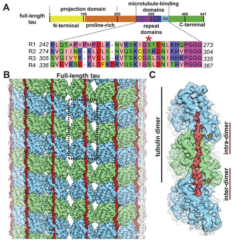
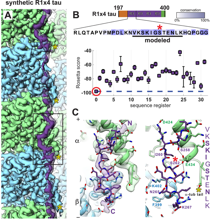
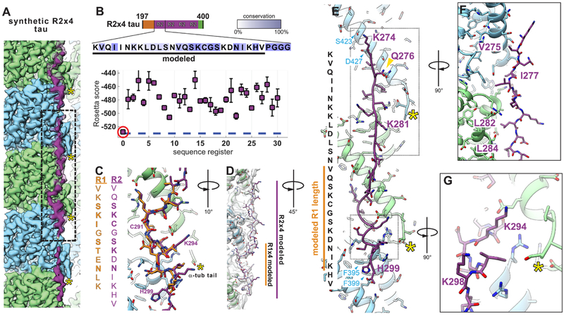
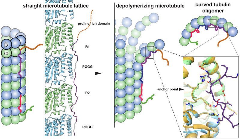
Tau is a developmentally regulated axonal protein that stabilizes and bundles microtubules (MTs). Its hyperphosphorylation is thought to cause detachment from MTs and subsequent aggregation into fibrils implicated in Alzheimer's disease. It is unclear which tau residues are crucial for tau-MT interactions, where tau binds on MTs, and how it stabilizes them. We used cryo-electron microscopy to visualize different tau constructs on MTs and computational approaches to generate atomic models of tau-tubulin interactions. The conserved tubulin-binding repeats within tau adopt similar extended structures along the crest of the protofilament, stabilizing the interface between tubulin dimers. Our structures explain the effect of phosphorylation on MT affinity and lead to a model of tau repeats binding in tandem along protofilaments, tethering together tubulin dimers and stabilizing polymerization interfaces.


Figures

Fig. 1.
Fig. 1.. Tau binding to microtubules.
(A) Schematic of tau domain architecture and assigned functions. The MT-binding domain of four repeats is defined as residues 242–367. Inset shows the sequence alignment of the four repeat sequences, R1–4, that make up the repeat domain. Ser262 is marked by the red asterisk. (B) Cryo-EM density map (4.1 Å overall resolution) of a MT decorated with full-length tau. Tau (red) appears as a nearly continuous stretch of density along PFs (α-tubulin in green, β-tubulin in blue). (C) The footprint of a continuous stretch of tau spans over three tubulin monomers, binding both across intra- and inter-dimer tubulin interfaces (only one repeat of tau is shown for clarity). Position of the C-termini of tubulin indicated with yellow asterisks.
Fig. 2.
Fig. 2.. Near-atomic resolution reconstruction of synthetic R1×4 tau on microtubules.
(A) In the 3.2 Å cryo-EM reconstruction of tau-bound MTs, the best-ordered segment of tau is bound at the interface between tubulin dimers (boxed) (lower threshold density for tau is shown in transparency). (B) Rosetta modeling reveals a single energetically preferred sequence register (red circle) for the best-ordered tau region, corresponding to a conserved (underlined) 12-residue stretch of residues within the R1 repeat sequence. All Rosetta simulations were repeated until convergence (100 models per register). Intensity of colored boxes indicates extent of conservation among tau homologs. S262 is indicated with red asterisk in (B) and (C). (C) Atomic model of tau and tubulin, with (left) and without (right) the density map, showing the interactions over the inter-dimer region. C-termini of tubulin indicated with yellow asterisks in (A) and (C).
Fig. 3.
Fig. 3.. High-resolution reconstruction of synthetic R2×4 tau on microtubules.
(A) The (3.9 Å) cryo-EM reconstruction of R2×4 is highly similar to that of R1×4, but reveals a longer stretch of ordered density for tau along the MT surface (lower threshold density for tau is shown in transparency). (B) Rosetta modeling supports a sequence register (red circle) for the R2 sequence binding to tubulin equivalent to that for R1 and shown in Fig. 2. All Rosetta simulations were repeated until convergence (100 models per register). (C) Major tau-tubulin interactions at the inter-dimer cleft are highly similar between the R1 (shown in goldenrod for easier visualization) and R2 (purple) sequences. (D) Extending the model to account for the additional density reveals an almost entire repeat of tau, spanning three tubulin monomers (centered on α-tubulin and contacting β- tubulin on either side) with an overall length of ~80 Å (the approximate length of a tubulin dimer). C-termini of tubulin indicated with yellow asterisks in (A), (C), (E), and (G). (E) Atomic model of the R2 repeat, shown along with the corresponding R2 sequence. The position of the previously studied gold-labeled residue in MAP2 at the equivalent position based on homology (16) is indicated with a gold arrowhead and is in very good agreement with our model (see also fig. S6). Boxed out regions show: (F) the hydrophobic packing of tau residues on the MT surface (see also fig. S8), and (G) the positioning of two R2 lysines with potential interactions with the α- tubulin acidic C-terminal tail.
Fig. 4.
Fig. 4.. Model of full-length tau binding to microtubules and tubulin oligomers.
Our structural data leads to a model of tau interaction with MTs in which the four repeats bind in tandem along a PF. Notice that we do not observe strong density for the region that would correspond to the PGGG motif, which is modeled in gray for illustrative purposes and must be highly flexible. Tau binding at the inter-dimer interface, interacting with both α- and β-tubulin, promotes association between tubulin dimers. The tau binding site is also the location of the previously identified “anchor point” [right-most box, gold is bent tubulin, PDB code 4I4T (31), and blue/green is straight tubulin, PDB code 3JAR (28)], and thus tau-tubulin interactions are unlikely to change significantly with protofilament peeling during disassembly (center) or when bound to small, curved tubulin oligomers (top right).

References

    1. Conde C, Cáceres A, Microtubule assembly, organization and dynamics in axons and dendrites. Nat. Rev. Neurosci 10, 319–332 (2009). doi:10.1038/nrn2631 Medline - DOI - PubMed
    1. Kosik KS, Orecchio LD, Bakalis S, Neve RL, Developmentally regulated expression of specific tau sequences. Neuron 2, 1389–1397 (1989). doi:10.1016/0896-6273(89)90077-9 Medline - DOI - PubMed
    1. Himmler A, Drechsel D, Kirschner MW, Martin DW Jr., Tau consists of a set of proteins with repeated C-terminal microtubule-binding domains and variable N-terminal domains. Mol. Cell. Biol 9, 1381–1388 (1989). doi:10.1128/MCB.9.4.1381 Medline - DOI - PMC - PubMed
    1. Ennulat DJ, Liem RK, Hashim GA, Shelanski ML, Two separate 18-amino acid domains of tau promote the polymerization of tubulin. J. Biol. Chem 264, 5327–5330 (1989). Medline - PubMed
    1. Butner KA, Kirschner MW, Tau protein binds to microtubules through a flexible array of distributed weak sites. J. Cell Biol 115, 717–730 (1991). doi:10.1083/jcb.115.3.717 Medline - DOI - PMC - PubMed

Publication types