

The Effect of Fucoidan from the Brown Alga Fucus evanescence on the Activity of α- N-Acetylgalactosaminidase of Human Colon Carcinoma Cells
- PMID: 29748462
- PMCID: PMC5983286
- DOI: 10.3390/md16050155
The Effect of Fucoidan from the Brown Alga Fucus evanescence on the Activity of α- N-Acetylgalactosaminidase of Human Colon Carcinoma Cells
Abstract
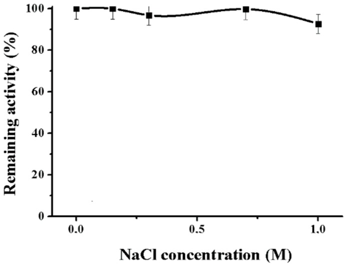
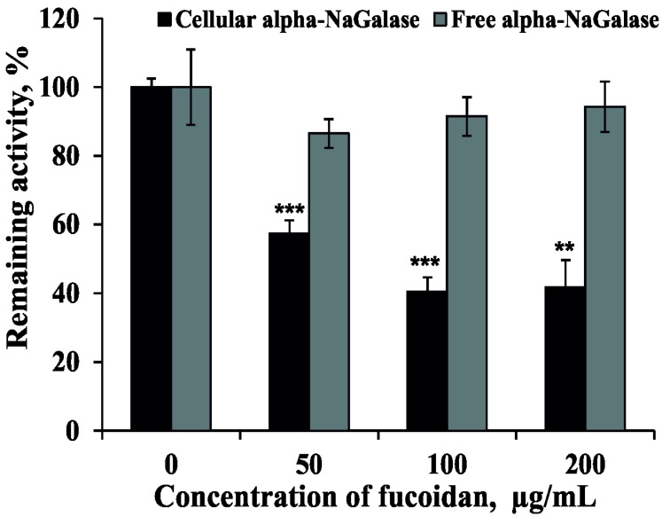
α-N-acetylgalactosaminidase (EC 3.2.1.49) (alpha-NaGalase) catalyzes the hydrolysis of N-acetamido-2-deoxy-α-d-galactoside residues from non-reducing ends of various complex carbohydrates and glycoconjugates. It is known that human cancer cells express an alpha-NaGalase, which accumulates in the blood plasma of patients. The enzyme deglycosylates the Gc protein-derived macrophage activating factor (GcMAF) and inhibits macrophage activity acting as an immunosuppressor. The high specific activity 0.033 ± 0.002 μmol mg−1 min−1 of the enzyme was found in human colon carcinoma cells DLD-1. The alpha-NaGalase of DLD-1 cells was isolated and biochemical characterized. The enzyme exhibits maximum activity at pH 5.2 and temperature 55 °C. The Km is 2.15 mM, Vmax⁻0.021 μmol min−1 mL−1, kcat⁻1.55 min−1 and kcat/Km⁻0.72 min−1 mM−1 at 37 °C, pH 5.2. The effects of fucoidan from the brown alga Fucus evanescence on the activity of alpha-NaGalase in human colon carcinoma DLD-1 cells and on the biosynthesis of this enzyme were investigated. It was shown that fucoidan did not inhibit free alpha-NaGalase, however, it reduced the expression of the enzyme in the DLD-1 cells at IC50 73 ± 4 μg mL−1.
Keywords: DLD-1; Fucus evanescence; alpha-NaGalase; brown alga; colon carcinoma cells; fucoidan; macrophage activating factor; α-N-acetylgalactosaminidase.
Conflict of interest statement
The authors declare no conflict of interest.
Figures


Similar articles
-
Immunotherapy of metastatic breast cancer patients with vitamin D-binding protein-derived macrophage activating factor (GcMAF).Int J Cancer. 2008 Jan 15;122(2):461-7. doi: 10.1002/ijc.23107. Int J Cancer. 2008. Retraction in: Int J Cancer. 2014 Sep 15;135(6):1509. PMID: 17935130 Retracted.
-
Tumor cell alpha-N-acetylgalactosaminidase activity and its involvement in GcMAF-related macrophage activation.Comp Biochem Physiol A Mol Integr Physiol. 2002 May;132(1):1-8. doi: 10.1016/s1095-6433(01)00522-0. Comp Biochem Physiol A Mol Integr Physiol. 2002. PMID: 12062184
-
Effect of salivary gland adenocarcinoma cell-derived alpha-N-acetylgalactosaminidase on the bioactivity of macrophage activating factor.Int J Oncol. 2004 Mar;24(3):521-8. Int J Oncol. 2004. PMID: 14767536
-
Is α-N-acetylgalactosaminidase the key to curing cancer? A mini-review and hypothesis.J BUON. 2017 Nov-Dec;22(6):1372-1377. J BUON. 2017. PMID: 29332325 Review.
-
Immunotherapy with GcMAF revisited - A critical overview of the research of Nobuto Yamamoto.Cancer Treat Res Commun. 2022;31:100537. doi: 10.1016/j.ctarc.2022.100537. Epub 2022 Feb 18. Cancer Treat Res Commun. 2022. PMID: 35217488 Review.
Cited by
-
Effects of Sponge-Derived Alkaloids on Activities of the Bacterial α-D-Galactosidase and Human Cancer Cell α-N-Acetylgalactosaminidase.Biomedicines. 2021 May 5;9(5):510. doi: 10.3390/biomedicines9050510. Biomedicines. 2021. PMID: 34063022 Free PMC article.
-
Effect of Phlorotannins from Brown Algae Costaria costata on α-N-Acetylgalactosaminidase Produced by Duodenal Adenocarcinoma and Melanoma Cells.Mar Drugs. 2022 Dec 30;21(1):33. doi: 10.3390/md21010033. Mar Drugs. 2022. PMID: 36662206 Free PMC article.
References
-
- Santos-Pereira J.M., Muñoz-Galván S. Biomarkers of colorectal cancer: A genome-wide perspective. Cancer Transl. Med. 2016;2:182–188.
MeSH terms
Substances
LinkOut - more resources
Full Text Sources
Other Literature Sources
Research Materials
Miscellaneous
