. 2018 May 10;9(1):1849.
doi: 10.1038/s41467-018-03917-2.

Genome-wide and high-density CRISPR-Cas9 screens identify point mutations in PARP1 causing PARP inhibitor resistance

Affiliations

Genome-wide and high-density CRISPR-Cas9 screens identify point mutations in PARP1 causing PARP inhibitor resistance

Stephen J Pettitt et al. Nat Commun. .

Abstract

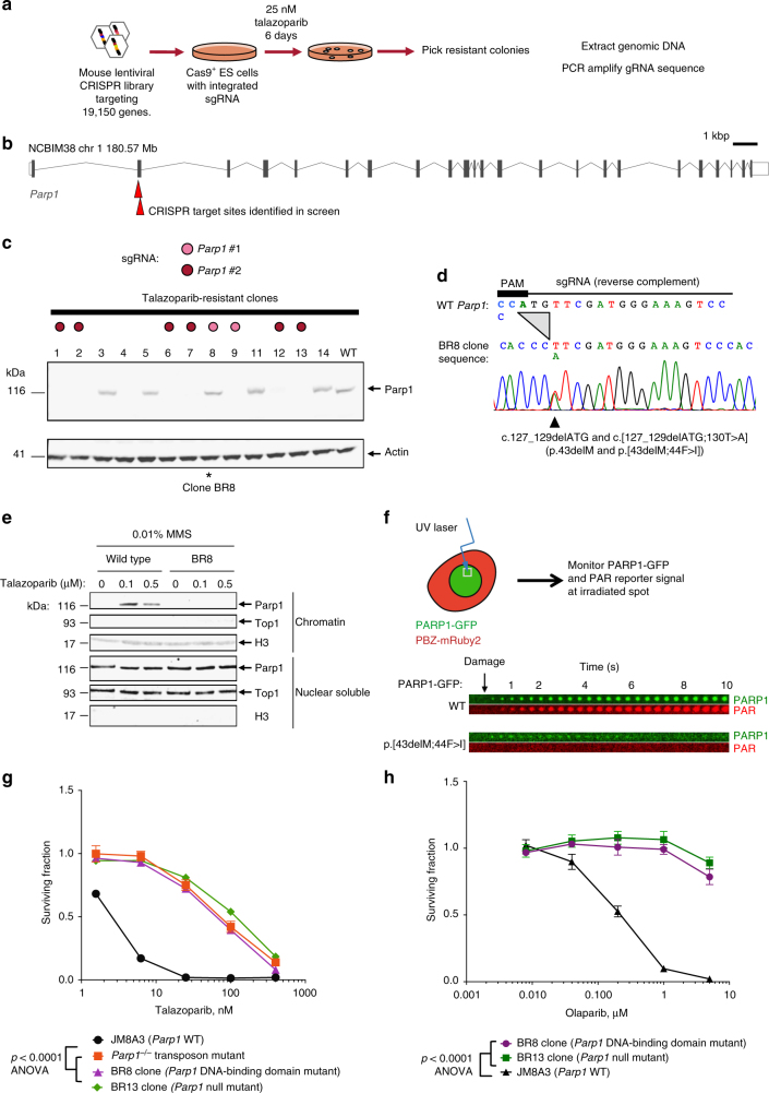
Although PARP inhibitors (PARPi) target homologous recombination defective tumours, drug resistance frequently emerges, often via poorly understood mechanisms. Here, using genome-wide and high-density CRISPR-Cas9 "tag-mutate-enrich" mutagenesis screens, we identify close to full-length mutant forms of PARP1 that cause in vitro and in vivo PARPi resistance. Mutations both within and outside of the PARP1 DNA-binding zinc-finger domains cause PARPi resistance and alter PARP1 trapping, as does a PARP1 mutation found in a clinical case of PARPi resistance. This reinforces the importance of trapped PARP1 as a cytotoxic DNA lesion and suggests that PARP1 intramolecular interactions might influence PARPi-mediated cytotoxicity. PARP1 mutations are also tolerated in cells with a pathogenic BRCA1 mutation where they result in distinct sensitivities to chemotherapeutic drugs compared to other mechanisms of PARPi resistance (BRCA1 reversion, 53BP1, REV7 (MAD2L2) mutation), suggesting that the underlying mechanism of PARPi resistance that emerges could influence the success of subsequent therapies.


Conflict of interest statement

C.J.L. and A.A. are named inventors on patents describing the use of PARP inhibitors and stand to gain from their development as part of the ICR “Rewards to Inventors” scheme. The remaining authors declare no competing interests.

Figures

Fig. 1
Fig. 1
A genome-wide CRISPR screen for PARP inhibitor resistance identifies in-frame Parp1 mutants. a Experimental scheme. b Locations of Parp1 guide RNA target sites in exon two of the mouse Parp1 gene. c Parp1 western blot of lysates from talazoparib-resistant clones identified in the CRISPR screen. Individual clones are colour-coded according to sgRNA present (see key). Clones 1, 2, 6, 7, 9, 12 and 13 with Parp1 sgRNAs have lost Parp1 protein expression, whilst Parp1 sgRNA clone 8 (BR8, *) has retained Parp1 expression. d Clone BR8 has an in-frame Parp1 deletion and a Parp1 substitution mutation. Sanger sequencing trace of the Parp1 sgRNA target site is shown, illustrating a 3 bp deletion on both alleles and a heterozygous c.130T>A substitution mutation (p.44F>I) close to the CRISPR PAM site. e Parp1 is not trapped in the chromatin fraction by PARP inhibitor in the BR8 clone. Western blots illustrating Parp1 in the chromatin and nuclear soluble fractions of wild-type ES cells and Parp1 mutant BR8 cells exposed to talazoparib. Data shown are representative of two experiments. f PARP1 protein with a p.[43delM;44F>I] mutation has impaired recruitment to damaged DNA and does not initiate PAR synthesis at damaged DNA. Localisation of PARP1-GFP to damaged DNA was estimated by visualising GFP signal at the microirradiated spot, as was the generation of PAR at the damaged site by use of a PAR-binding PBZ-mRuby2 probe (see schematic). The time course of PARP1-GFP and PBZ-mRuby2 signals from CAL51 PARP1–/– cells transfected with PARP1-GFP (top) and PARP1-p.[43delM;44F>I]-GFP (bottom) are shown. “Damage” denotes time at which microirradiation was carried out. g, h Dose response curves illustrating that clone BR8 is resistant to both talazoparib (g) and olaparib (h). BR13 is an ES cell mutant with no Parp1 protein expression (see panel c); a transposon-mutagenised, Parp1 null mutant is also shown (red). Clone BR8 vs. BR13 and Parp1–/– transposon, p = ns, ANOVA. Parp1 mutant clones vs. wild-type cells, p < 0.0001, ANOVA. Mean of five replicates plotted, error bars show SD. Surviving fractions were calculated relative to DMSO-exposed cells for each mutant
Fig. 2
Fig. 2
A focused CRISPR-Cas9 mutagenesis “tag, mutate and enrich” screen identifies PARP1 mutations outside the DNA-binding domain that cause PARP inhibitor resistance. a Experimental “tag, mutate and enrich” scheme to isolate missense and in-frame PARP1 mutants associated with PARP inhibitor resistance. b Translated alignment of PARP1 amino-acid mutations identified in talazoparib-resistant cells isolated from lentiviral sgRNA pool 1 (designed to target ZnF1 and ZnF2 domains) from the screen shown in a. By comparing the multiple different mutations isolated from pool 1, a PARP1 p.119_120delKS minimal mutation associated with resistance was identified. Some insertions and larger deletions are omitted for clarity (see Supplementary Data 2). c PARP1 p.119_120delKS mutation abolishes recruitment of a PARP1-GFP fusion to sites of microirradiated DNA damage. CAL51 PARP1–/– cells were transfected with a wild-type PARP1-GFP cDNA construct or a PARP1 p.119_120delKS-GFP fusion cDNA expression construct and then exposed to localised ionising radiation (a microirradiated spot) as in f. Time course of PARP1-GFP signal at microirradiated site is shown. d Location of three mutations associated with PARPi resistance on a model of the PARP1 DNA structure. e PARP1-N329Q and HD742F mutations ablate PARP1 trapping, while 848delY partially reduces trapping. Western blot from PARP1 trapping assay for three PARP1-GFP mutants and wild-type PARP1-GFP is shown. MMS-treated cells were lysed and fractionated into nuclear soluble (NS) and chromatin (C) fractions as described in Methods. Blot was probed with an anti-GFP antibody. f PARP1-848delY mutation alters PARP1 localisation kinetics at sites of DNA damage. Microirradiation and PAR synthesis phenotypes of N329Q and 848delY PARP1 mutants. Green—PARP1-GFP signal, red—PAR sensor (PBZ-mRuby2). Note lower recruitment relative to wild type of both mutants, but retention of PAR synthesis in 848delY. g Model of intramolecular communication between the DNA-binding ZnF1 domain and the catalytic (CAT) domain based on mutants identified from screens. (1) 45delD mutation in first zinc finger, (2) HD742F mutation shown in d, (3) E688 (Supplementary Data 2)
Fig. 3
Fig. 3
PARP1 mediates PARPi sensitivity in BRCA1 mutant cell lines. a Cas9-expressing BRCA1 mutant SUM149 cells were transduced with PARP1-ZnF or hsvTK (non-targeting control) lentiviral sgRNA particles, selected in puromycin and exposed to the indicated talazoparib concentrations for 7 days. Representative SRB-stained 24-well plate image is shown. b Western blot showing PARP1 expression in talazoparib-resistant SUM149 clones TR1 and TR2. Par, parental SUM149 cells. TR2 has an in-frame PARP1-p.43_45delMFD mutation. c SUM149 clones TR1 and TR2 are highly talazoparib-resistant. Plot shows survival relative to DMSO-exposed cells after exposure to the indicated concentration of talazoparib for 7 days. Mean of five replicates plotted, error bars show SD. d PARP1 mutant SUM149-TR2 xenografts do not respond to talazoparib treatment, whereas PARP1 wild-type SUM149 xenografts do. Top panels show Kaplan–Meier survival curves, with a tumour volume of 1000 mm3 (after which mice were sacrificed) used as a surrogate end point. Lower panels show volume of individual tumours, normalised according to tumour volume on the day of randomisation (indicated by the arrow). e Pools of PARP1 guides cause talazoparib resistance in the BRCA1 mutant ovarian cancer cell line COV362. Cas9-expressing COV362 cells were transduced with the indicated lentiviral guide pool, selected in puromycin and assayed as in c. f Silencing of residual BRCA1 function, or BRCA2, induces synthetic lethality with PARP1 genetic loss in SUM149 cells. B1.S* is a SUM149 derivative with a secondary BRCA1 mutation that confers PARP inhibitor resistance. SRB-stained 24-well plate image is shown, 7 days after siRNA transfection. g Colony forming assay using SUM149-Cas9 parental cells and the talazoparib-resistant daughter clone, TR2. Cells were transfected as in f, plated on 6-well plates and colonies stained and counted 2 weeks later. siRNA targeting BRCA1 or BRCA2 significantly reduces survival in PARP1 mutant cells (TR2) compared to the parental SUM149 line (t-test). h BRCA1 and BRCA2 silencing is also synthetically lethal in COV362 PARP1 mutant clones A4 (p.119_120delKS) and D1 (p.848delY/YK). Colony formation assay as in g, p values for t-test shown. Mean of three replicates plotted, error bars show SD
Fig. 4
Fig. 4
A high-density tiling CRISPR-Cas9 screen enables functional annotation of a PARP1 mutation observed in an olaparib-resistant patient. a Positions of sgRNA target sites in the dense PARP1 tiling library mapped onto the PARP1 coding DNA sequence shown in b. b Positions of mutations identified in the dense PARP1 tiling screen. Number of mutant reads for in-frame mutant alleles affecting each base is shown on the y-axis normalised to per-base coverage (mutant reads/total coverage × 1000). Only sites with coverage >500 reads are shown. The experiment was repeated in duplicate from two independently tagged SUM149 PARP1-GFP cell lines (clone 5 and 8, shown in red and blue). Arrows highlight the mutations observed at inter-domain contacts as shown in e. c Positions of protein domains mapped onto the PARP1 protein sequence. Red triangles indicate DNA–protein contacts based on crystal structures, blue circles show the location of the patient mutation identified in this study. Grey arcs represent key inter-domain contacts as shown in e and Fig. 2g. d Ribbon plot of ZnF1 and ZnF2 bound to a single-strand break (PDB: 2N8A). Ribbons are coloured red by residue based on the frequency of mutations observed in the tiling screen affecting that residue as shown in b. The thickness of the ribbon is proportional to the frequency of mutations observed affecting that residue. e Ribbon plot of the PARP1-DSB crystal structure (PDB: 4OQB) highlighting the clustering of mutations along the hydrogen-bonding axis postulated in Fig. 2g and the A925 residue that abuts Y848 (inset 2). Regions marked 1 and 2 are magnified. f Analysis of trapping for the R591C patient mutation by microirradiation with or without talazoparib as shown. Although the R591C mutant can bind DNA at sites of damage (blue line), it is not trapped in the presence of talazoparib (purple line)

References

    1. Farmer H, et al. Targeting the DNA repair defect in BRCA mutant cells as a therapeutic strategy. Nature. 2005;434:917–921. doi: 10.1038/nature03445. - DOI - PubMed
    1. Bryant HE, et al. Specific killing of BRCA2-deficient tumours with inhibitors of poly(ADP-ribose) polymerase. Nature. 2005;434:913–917. doi: 10.1038/nature03443. - DOI - PubMed
    1. Lord CJ, Ashworth A. PARP inhibitors: synthetic lethality in the clinic. Science. 2017;355:1152–1158. doi: 10.1126/science.aam7344. - DOI - PMC - PubMed
    1. Mortusewicz O, Amé JC, Schreiber V, Leonhardt H. Feedback-regulated poly(ADP-ribosyl)ation by PARP-1 is required for rapid response to DNA damage in living cells. Nucleic Acids Res. 2007;35:7665–7675. doi: 10.1093/nar/gkm933. - DOI - PMC - PubMed
    1. Murai J, et al. Stereospecific PARP trapping by BMN 673 and comparison with olaparib and rucaparib. Mol. Cancer Ther. 2014;13:433–443. doi: 10.1158/1535-7163.MCT-13-0803. - DOI - PMC - PubMed

Publication types

MeSH terms