. 2018 May 10;8(1):7479.
doi: 10.1038/s41598-018-25840-8.

Informative three-dimensional survey of cell/tissue architectures in thick paraffin sections by simple low-vacuum scanning electron microscopy

Affiliations

Informative three-dimensional survey of cell/tissue architectures in thick paraffin sections by simple low-vacuum scanning electron microscopy

Akira Sawaguchi et al. Sci Rep. .

Abstract

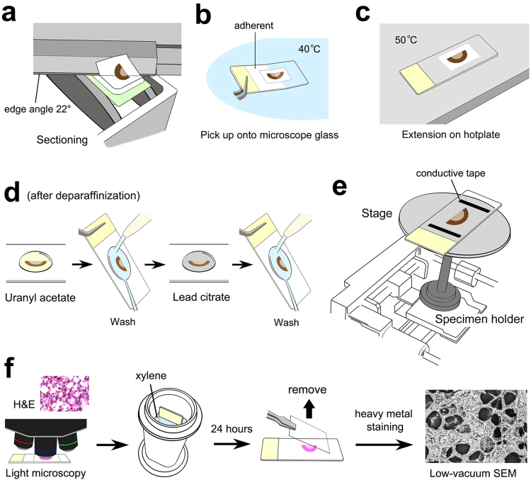
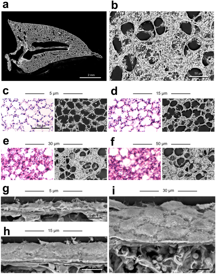
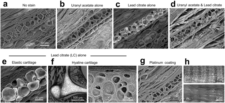
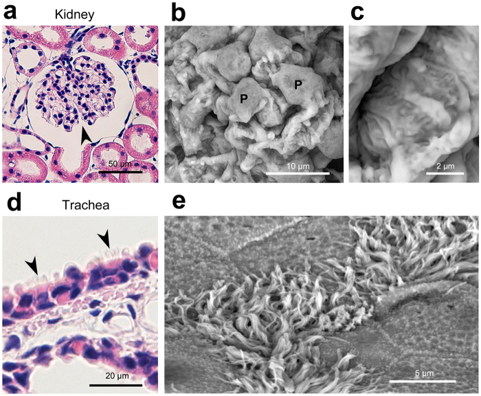
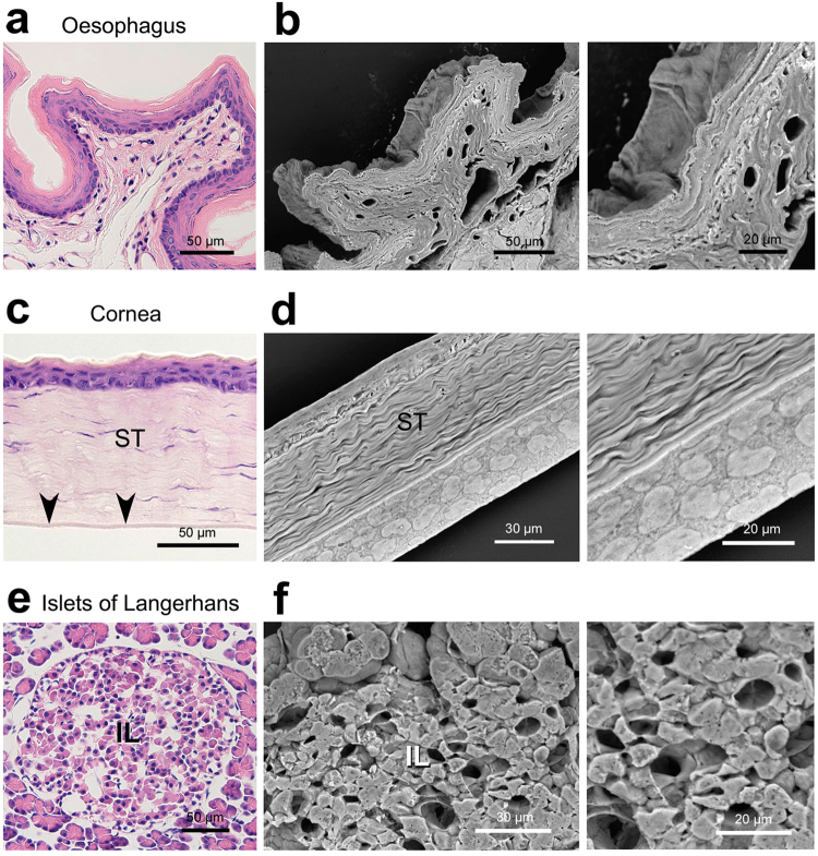
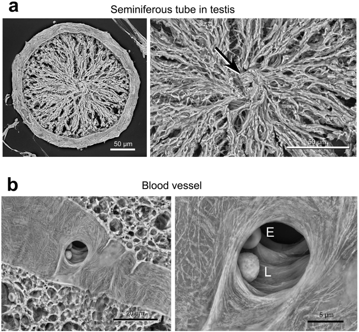
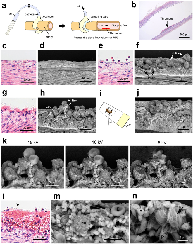
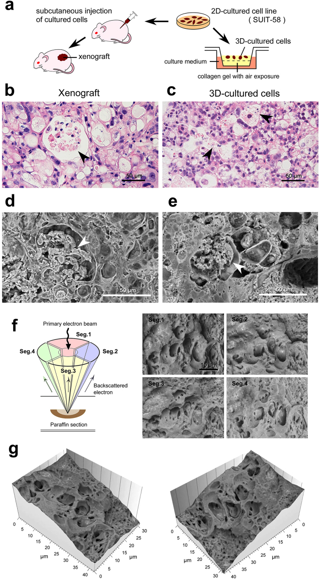
Recent advances in bio-medical research, such as the production of regenerative organs from stem cells, require three-dimensional analysis of cell/tissue architectures. High-resolution imaging by electron microscopy is the best way to elucidate complex cell/tissue architectures, but the conventional method requires a skillful and time-consuming preparation. The present study developed a three-dimensional survey method for assessing cell/tissue architectures in 30-µm-thick paraffin sections by taking advantage of backscattered electron imaging in a low-vacuum scanning electron microscope. As a result, in the kidney, the podocytes and their processes were clearly observed to cover the glomerulus. The 30 µm thickness facilitated an investigation on face-side (instead of sectioned) images of the epithelium and endothelium, which are rarely seen within conventional thin sections. In the testis, differentiated spermatozoa were three-dimensionally assembled in the middle of the seminiferous tubule. Further application to vascular-injury thrombus formation revealed the distinctive networks of fibrin fibres and platelets, capturing the erythrocytes into the thrombus. The four-segmented BSE detector provided topographic bird's-eye images that allowed a three-dimensional understanding of the cell/tissue architectures at the electron-microscopic level. Here, we describe the precise procedures of this imaging method and provide representative electron micrographs of normal rat organs, experimental thrombus formation, and three-dimensionally cultured tumour cells.


Conflict of interest statement

The authors declare no competing interests.

Figures

Figure 1
Figure 1
Sample preparation and anatomical findings by Thick PS-LvSEM. (ae) Illustration of the basic workflow. (a) Sectioning. (b) Mounting onto adherent coating microscope slides. (c) Extension on a hot plate. (d) Staining with uranyl acetate followed by Reynold’s lead citrate. (e) Setting onto the wide stage of a specimen holder. (f) Illustrated procedure for the correlative light and electron microscopy.
Figure 2
Figure 2
Preliminary examinations to determine the deal thickness of paraffin sections. (a,b) Lung. Montage image of whole section (a) and high-power view of the pulmonary alveoli with its wall face image (b). (cf) Correlative light (H&E staining) and Thick PS-LvSEM microscopy of rat lung paraffin sections in a series of 5, 15, 30, and 50 µm thickness. The sectioned features of alveolar septum are dominant in the 5 µm (c) and 15 µm (d) sections. On the other hand, the wall-face features occupy more than half the area in the 30 µm (e) and 50 µm (f) sections. Note the unavoidable cracks in the 50 µm sections. (gi) Comparison of oblique-section images of pulmonary pleura in 5 µm (g), 15 µm (h), and 30 µm (i) sections.
Figure 3
Figure 3
Preliminary examinations to determine the stainability of 30 µm paraffin sections. (ae,g,h) Rat auricle and (f) larynx. (a) No stain. (b) 1% uranyl acetate in 70% methanol (UA) alone. (c) Reynolds’ lead citrate solution (LC) alone. (d) UA and LC. (e,f) High-power view of elastic cartilage (e), and hyaline cartilage (f) in larynx stained with LC alone. Note the remarkable LC-stainability of the cartilage, as shown in the high-power view of hyaline matrix (right in f). (g) Platinum coating. (h) Comparison between UA/LC staining (upper) and platinum coating (lower) on skeletal muscle fibre. The skeletal muscle striation is significantly masked by the platinum coating.
Figure 4
Figure 4
Representative micrographs of rat kidney and trachea. Electron micrographs were taken from 30 µm sections. (ac) Kidney. (a) Overview of the glomerulus (arrowhead) within the renal corpuscle. (b) Podocytes (P) covering the glomerular capillaries. (c) High-power view demonstrates the engaged processes. (d,e) Trachea. Note the ciliated epithelial cells (arrowheads) distinguished by Thick PS-LvSEM.
Figure 5
Figure 5
Representative micrographs of rat oesophagus, cornea, and pancreas. Electron micrographs were taken from 30 µm sections. (a,b) Oesophagus. Thick PS-LvSEM demonstrates the multiple layers of stratified epithelium. (c,d) Cornea. The face image of endothelium, rarely seen in 5 µm sections (arrowheads in c), is clearly shown in the 30 µm section. Note the collagen fibres in its stroma (ST). (e,f) Islets of Langerhans (IL) in the pancreas. The capillary network is seen, a characteristic of endocrine organs.
Figure 6
Figure 6
Representative micrograph of rat testis and blood vessel. Electron micrographs were taken from 30 µm sections. (a) Seminiferous tubule in the testis. Differentiated sperm cells are assembled in the centre (arrow). (b) A blood vessel in the spinal cord. Note the vascular endothelium and its bifurcation with erythrocyte (E) and leukocyte (L).
Figure 7
Figure 7
Application of Thick PS-LvSEM to pathological experiments. (an) An experimental model of thrombus formation induced by disturbing the blood flow in the rabbit femoral artery. All images were taken from 30 µm sections, except the light micrographs from 5 µm sections with H&E staining (b,c,e,g,l). (a) Illustration of the arterial blood flow disturbance. (b) Overview of the mural thrombus formation. (c,d) Normal endothelium. (e,f) Adhesion of leukocytes (Leu) and platelets (PLT) on the erosive endothelium. (gk) Aggregations of leukocytes (Leu), erythrocytes (Ery), and platelets, attaching to the sub-endothelium exposed in close proximity to the thrombus. (i) Illustrated use of the optional 45° tilt-holder. (j) Tilted top view of the aggregation. (k) Comparison of accelerating voltages. Note the higher signal-to-noise ratio at 15 kV, and the surface configuration revealed at 5 kV. (ln) Core portion of the mural thrombus consisting of erythrocytes (E), leukocytes (Leu), platelets (P, and arrowhead in l), and fibrin fibres (arrow). (n) Note the network of fibrin fibres and platelets capturing the erythrocytes.
Figure 8
Figure 8
Application of Thick PS-LvSEM to oncological experiments using SUIT-58 cell line. (a) Illustration of the comparative experiments between xenograft and three-dimensionally cultured cells. (b,c) Light micrographs of H&E stained 5 µm sections and (dg) Thick PS-LvSEM micrographs of 30 µm sections. Note the similar profiles of vacuole formation enclosing necrotic cell debris (arrowheads). (f) Illustration of the four-segmented BSE detectors (left) and collected micrographs by each detector (right). (g) Topographic images reconstructed from the raw micrographs of the four-segmented BSE images.

References

    1. Shi Y, Inoue H, Wu JC, Yamanaka S. Induced pluripotent stem cell technology: a decade of progress. Nat. Rev. Drug Discov. 2017;16:115–130. doi: 10.1038/nrd.2016.245. - DOI - PMC - PubMed
    1. Stoltz JF, Zhang L, Ye JS, De Isla N. Organ reconstruction: Dream or reality for the future. Biomed Mater. Eng. 2017;28:S121–S127. - PubMed
    1. Chen S, et al. Genome-wide CRISPR screen in a mouse model of tumor growth and metastasis. Cell. 2015;160:1246–1260. doi: 10.1016/j.cell.2015.02.038. - DOI - PMC - PubMed
    1. Talbot MJ, White RG. Cell surface and cell outline imaging in plant tissues using the backscattered electron detector in a variable pressure scanning electron microscope. Plant Methods. 2013;9:40. doi: 10.1186/1746-4811-9-40. - DOI - PMC - PubMed
    1. Tardi NJ, Cook ME, Edwards KA. Rapid phenotypic analysis of uncoated Drosophila samples with low-vacuum scanning electron microscopy. Fly. 2012;6:184–192. doi: 10.4161/fly.20525. - DOI - PubMed