. 2018 May 1;9(5):555.
doi: 10.1038/s41419-018-0619-5.

Gene-by-environment interactions that disrupt mitochondrial homeostasis cause neurodegeneration in C. elegans Parkinson's models

Affiliations

Gene-by-environment interactions that disrupt mitochondrial homeostasis cause neurodegeneration in C. elegans Parkinson's models

Hanna Kim et al. Cell Death Dis. .

Abstract

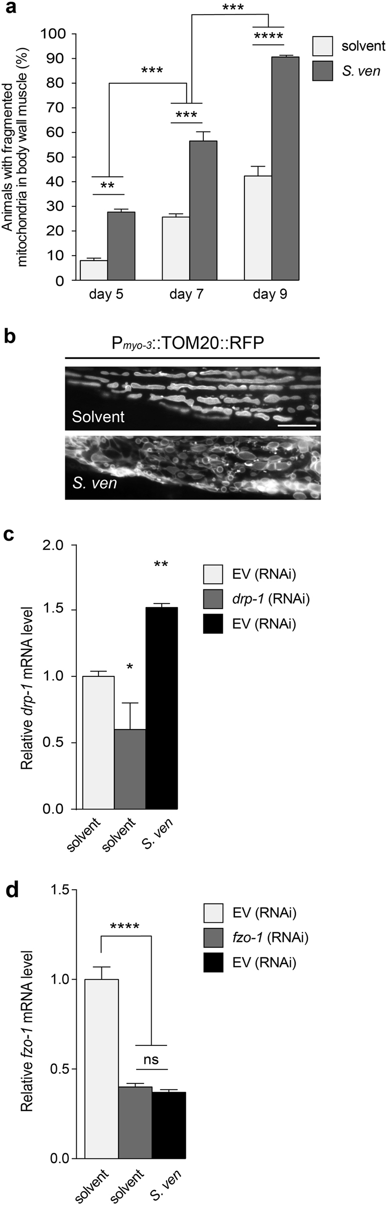
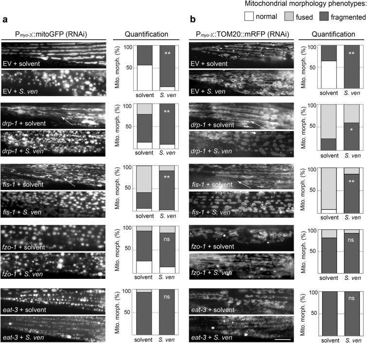
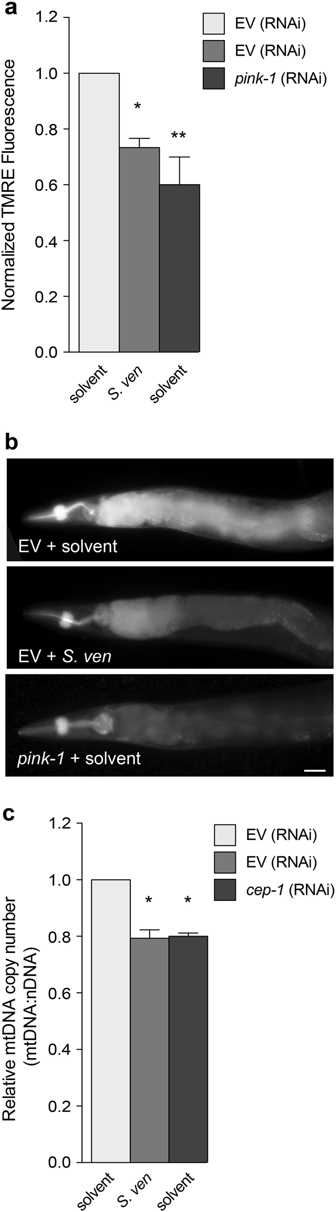
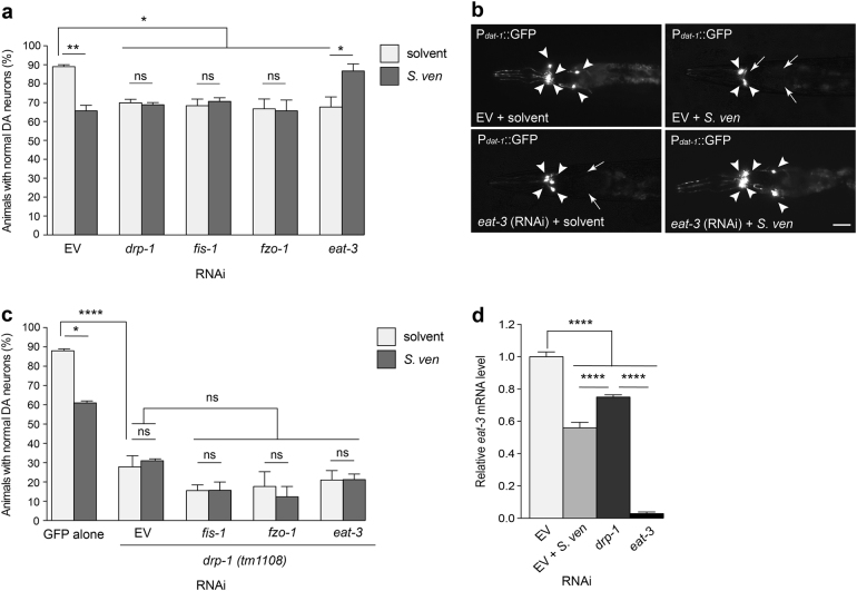
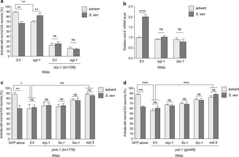
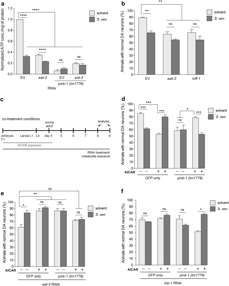
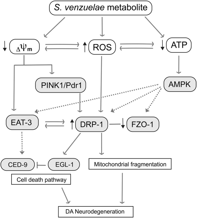
Parkinson's disease (PD) is a complex multifactorial disorder where environmental factors interact with genetic susceptibility. Accumulating evidence suggests that mitochondria have a central role in the progression of neurodegeneration in sporadic and/or genetic forms of PD. We previously reported that exposure to a secondary metabolite from the soil bacterium, Streptomyces venezuelae, results in age- and dose-dependent dopaminergic (DA) neurodegeneration in Caenorhabditis elegans and human SH-SY5Y neurons. Initial characterization of this environmental factor indicated that neurodegeneration occurs through a combination of oxidative stress, mitochondrial complex I impairment, and proteostatic disruption. Here we present extended evidence to elucidate the interaction between this bacterial metabolite and mitochondrial dysfunction in the development of DA neurodegeneration. We demonstrate that it causes a time-dependent increase in mitochondrial fragmentation through concomitant changes in the gene expression of mitochondrial fission and fusion components. In particular, the outer mitochondrial membrane fission and fusion genes, drp-1 (a dynamin-related GTPase) and fzo-1 (a mitofusin homolog), are up- and down-regulated, respectively. Additionally, eat-3, an inner mitochondrial membrane fusion component, an OPA1 homolog, is also down regulated. These changes are associated with a metabolite-induced decline in mitochondrial membrane potential and enhanced DA neurodegeneration that is dependent on PINK-1 function. Genetic analysis also indicates an association between the cell death pathway and drp-1 following S. ven exposure. Metabolite-induced neurotoxicity can be suppressed by DA-neuron-specific RNAi knockdown of eat-3. AMPK activation by 5-amino-4-imidazole carboxamide riboside (AICAR) ameliorated metabolite- or PINK-1-induced neurotoxicity; however, it enhanced neurotoxicity under normal conditions. These studies underscore the critical role of mitochondrial dynamics in DA neurodegeneration. Moreover, given the largely undefined environmental components of PD etiology, these results highlight a response to an environmental factor that defines distinct mechanisms underlying a potential contributor to the progressive DA neurodegeneration observed in PD.


Conflict of interest statement

The authors declare that they have no conflict of interest.

Figures

Fig. 1
Fig. 1. The S. venezuelae (S. ven) metabolite causes perturbations of mitochondrial fission and fusion, resulting in time-dependent mitochondrial fragmentation in C. elegans.
a Quantification of mitochondrial fragmentation as indicated at young adult (5-day old), middle (7-day old), and older aged (9-day old) nematodes in response to metabolite exposure using Pmyo-3:TOM20:mRFP. S. ven metabolite significantly enhanced mitochondrial fragmentation in aging nematodes. Data represented as mean ± S.E.M.; n = 30 animals per replicate, three independent replicates; two-way ANOVA with Tukey’s post hoc test for multiple comparisons. **P< 0.01, ***P< 0.001, and ****P< 0.0001. b TOM20::mRFP images of the representative mitochondrial morphology following exposure of solvent or metabolite at day 5 post-hatching. The metabolite causes disordered and small donut-shaped morphology in the mitochondrial outer membrane. The scale bar is 20 μm. c, d Animals exposed to solvent (EtAc) or S. ven metabolite were assessed for drp-1 and fzo-1 mRNA levels by qRT-PCR. Metabolite exposure significantly increased drp-1 expression and reduced fzo-1 expression levels compared to empty vector (EV) solvent control. Relative mRNA expression levels were normalized to EV control. Data represented as mean ± S.E.M.; three replicates comprising at least 100 animals each; one-way ANOVA with Tukey’s post hoc test for multiple comparisons. *P< 0.05, **P< 0.01, and ****P< 0.0001.
Fig. 2
Fig. 2
The effect of S. ven metabolite-mediated fission and fusion on the mitochondrial matrix (a) and mitochondrial outer membrane (b) in C. elegans body-wall muscles. Transgenic nematodes expressing Pmyo-3::mitoGFP were analyzed to monitor mitochondrial matrix in response to each treatment, and mitochondrial outer membrane was detected with Pmyo-3::TOM20::mRFP strain. Representative mitoGFP and TOM20 images (left panel in a and b) and quantification of the different mitochondrial morphologies observed (right panel with graphical stacks in a and b, Y axis represents distribution of mitochondrial morphology phenotypes in worm populations in percentage) are shown. Compared with solvent treatment, populations of RNAi-treated fission gene-associated drp-1 and fis-1 animals exhibited significantly more mitochondrial fragmentation following metabolite exposure, characterized by disordered and small circularly shaped mitochondria at day 8 post-hatching. Animals reduced for the fusion regulators, fzo-1 and eat-3, displayed mitochondrial fragmentation following exposure to metabolite in a manner similar to solvent-treated animals, respectively. Arrows indicate representative areas of mitochondrial fusion. These data are presented as mean ± S.E.M.; n = 30 animals per replicate, two independent replicates; one-way ANOVA with Tukey’s post hoc test for multiple comparisons. *P< 0.05, **P< 0.01. The scale bar is 20 μm.
Fig. 3
Fig. 3. The effect of S. ven metabolite-mediated fission and fusion on mitochondrial function.
a Relative mitochondrial uptake of the fluorescent dye tetramethylrhodamine ethyl ester (TMRE) was assessed for the mitochondrial membrane potential (ΔΨm) of C. elegans. Animals (wild-type N2) exposed to S. venezuelae metabolite had a significantly lower ΔΨm then solvent-treated EV control animals at day 8 post-hatching. RNAi knockdown of pink-1 was used as a positive control. Relative TMRE fluorescent intensity was normalized to EV solvent control. These data are presented as mean ± S.E.M.; n = 30 animals per replicate, three independent replicates; one-way ANOVA with Dunnett’s post hoc test for multiple comparisons. *P< 0.05, **P< 0.01. b Representative images of TMRE-stained nematodes following treatment with solvent and metabolite, respectively. The scale bar is 5 μm. c Quantitation of mitochondrial DNA (mtDNA) copy number by qRT-PCR in N2 animals. The metabolite caused a relative decrease of mtDNA copy number in comparison to EV solvent control. cep-1 (RNAi) was included as a positive control. Data represented as mean ± S.E.M.; three biological and three technical replicates; one-way ANOVA with Tukey’s post hoc test for multiple comparisons. *P< 0.05
Fig. 4
Fig. 4. Down-regulation of eat-3 suppresses neurotoxicity caused by the metabolite.
a, b RNAi knockdown of fission (drp-1 and fis-1) and fusion (fzo-1 and eat-3) components with solvent caused DA neurodegeneration at day 9 post-hatching. With metabolite exposure, RNAi treated with drp-1, fis-1, or fzo-1 animals did not exhibit enhanced DA neurotoxicity compared to the corresponding solvent control. In contrast, a combination of S. ven metabolite and RNAi targeting eat-3 resulted in significant resistance to neurotoxicity. The scale bar is 10 μm. These data are presented as mean ± S.E.M.; n = 30 animals per replicate, three independent replicates; one-way ANOVA with Tukey’s post hoc test for multiple comparisons. *P< 0.05, **P< 0.01. Representative images of the six anterior dopaminergic neurons are shown in (b), with degenerating or missing neurons marked by arrows and normal neurons indicated by arrowheads. c An RNAi-sensitive strain with GFP expression in DA neurons was crossed to drp-1(tm1108) mutant animals. Homozygosity was confirmed by PCR. Loss-of-function drp-1(tm1108) exacerbated DA neurotoxicity compared with GFP only solvent control at day 9 post-hatching. RNAi knockdown of eat-3 did not result in resistance of neurotoxicity as observed in Fig. 4a. Data are represented as mean ± S.E.M; n = 30 animals per replicate, three independent replicates; one-way ANOVA with Tukey’s post hoc test for multiple comparisons. *P < 0.05, *P < 0.0001. d eat-3 mRNA levels were measured by qRT-PCR in animals treated with solvent (EtAc), S. ven metabolite, drp-1 (RNAi), or eat-3 (RNAi). The animals treated with metabolite showed significant lower eat-3 mRNA levels than when drp-1 was depleted by RNAi. Data represented as mean ± S.E.M.; three replicates comprises at least 100 animals each; one-way ANOVA with Tukey’s post hoc test for multiple comparisons. ****P< 0.0001.
Fig. 5
Fig. 5. Exposure to S. ven metabolite is associated with cell death.
a Reduction of egl-1 (RNAi) caused neurodegeneration in comparison with EV solvent control; however, addition of metabolite showed significant reduction of neurotoxicity. Under the drp-1(tm1108) mutant background, the animals exhibited severe neurodegeneration, and the addition of metabolite did not cause further neurodegeneration in EV solvent control or egl-1 RNAi. These data are presented as mean ± S.E.M.; n = 30 animals per replicate, three independent replicates; one-way ANOVA with Tukey’s post hoc test for multiple comparisons. **P< 0.01. b ced-9 mRNA levels were measured by qRT-PCR. Metabolite exposure resulted in a significant increase in ced-9 transcriptional activity compared to EV solvent control, however, there was no significant change in ced-9 expression level when egl-1 or drp-1 was depleted (RNAi). Data are represented as mean ± S.E.M; three replicates comprised of at least 100 animals each; one-way ANOVA with Tukey’s post hoc test for multiple comparisons. ****P< 0.0001. c, d A dopaminergic (DA) neuron RNAi-sensitive strain with GFP expression in DA neurons was crossed to strains harboring mutant alleles for pink-1(tm1779) or pdr-1(gk448). Homozygosity was confirmed by PCR. Loss of either pink-1(tm1779) (c) or pdr-1(gk448) (d) function enhanced DA neurodegeneration with or without metabolite compared to GFP only solvent control at day 9 post-hatching. RNAi knockdown of eat-3 resulted in significant resistance to both pink-1 (c) and pdr-1-induced (d) neurotoxicity. Data presented as mean ± S.E.M.; n = 30 animals per replicate, three independent replicates; one-way ANOVA with Tukey’s post hoc test for multiple comparisons. *P< 0.05, **P < 0.01, ***P < 0.001, ****P < 0.0001.
Fig. 6
Fig. 6. AICAR-mediated AMPK activation rescues metabolite-induced dopaminergic neurodegeneration.
a The effect of eat-3 RNAi on ATP levels in pink-1(tm1779) mutant animals after exposure of solvent (EtAc) or metabolite. ATP content was measured in young adult nematodes using a luciferase-based assay. Values are mean ± S.E.M.; n = 3 independent samples with 100 worms in each; one-way ANOVA with Tukey’s post hoc test for multiple comparisons. ****P< 0.0001. b Reduction of either aak-2 or mff-1 enhanced DA neurotoxicity with or without metabolite in comparison with EV solvent control. Data represented as mean ± S.E.M.; n = 30 animals per replicate, three independent replicates; one-way ANOVA with Tukey’s post hoc test for multiple comparisons. **P< 0.005. c A timeline representing an experimental paradigm depicting the relative length of S. ven metabolite exposure and AICAR treatment. The abbreviations L1–L4 are the larval stages of C. elegans, and the ‘young adult’ designations represent days post-hatching. F1 animals were treated with either RNAi and/or S. ven metabolite from hatching to the day of analysis; neurodegeneration assays were performed at day 9. Animals were treated with 1 mM AICAR from hatching until day 4. d Animals expressing only GFP in the DA neurons treated with AICAR (alone) displayed enhanced DA neurodegeneration compared to solvent controls; however, metabolite-induced neurotoxicity was reduced by AICAR treatment. Furthermore, AICAR rescued neurotoxicity caused by pink-1(tm1779) in comparison to non-AICAR treated (solvent) animals with pink-1 mutation. AICAR did not rescue DA neuron cell death from the combined stress of metabolite and pink-1. Data are presented as mean ± S.E.M.; n = 30 animals per replicate, three independent replicates; one-way ANOVA with Tukey’s post hoc test for multiple comparisons. *P< 0.05, ***P< 0.001. e AICAR-mediated AMPK activation was sufficient to protect neurodegeneration caused by eat-3 deficiency (RNAi) alone or with metabolite, however, it could not reduce neurotoxicity in the absence of pink-1 function (tm1779) and eat-3 RNAi together. Data represented as mean ± S.E.M.; n = 30 animals per replicate, three independent replicates; one-way ANOVA with Tukey’s post hoc test for multiple comparisons. *P< 0.05, **P< 0.01. f AICAR-mediated AMPK activation failed to attenuate neurotoxicity from drp-1 (RNAi) depletion in the GFP only or pink-1(tm1779) mutant background; however, AICAR suppressed neurotoxicity in the pink-1 mutant background in combination with metabolite exposure. These data are presented as mean ± S.E.M.; n = 30 animals per replicate, three independent replicates; one-way ANOVA with Tukey’s post hoc test for multiple comparisons. *P< 0.05.
Fig. 7
Fig. 7. Proposed mechanism of altered mitochondrial homeostasis caused by S.
ven metabolite-induced toxicity. This experimental model illustrates our current understanding of the effect of S. ven metabolite on mitochondrial homeostatic mechanisms in the development of neurodegeneration. metabolite-induced ROS gradually impairs mitochondria through mitochondrial complex I damage, energy deprivation, and eventually, the loss of ΔΨm. As ΔΨm is decreased, C. elegans PINK-1 and PDR-1 would be mobilized to the OMM, which would then recruit the OMM fission factor, DRP-1, to the OMM to degrade the damaged organelle, resulting in mitochondrial fragmentation. Moreover, proteolytic cleavage of EAT-3 by the decline in ΔΨm and increased DRP-1 could lead to neuron cell death. In our genetic studies, the reduction of eat-3 (RNAi) attenuated neurotoxicity induced by metabolite or in the pink-1 mutant background, demonstrating that eat-3 depletion (RNAi) might counteract cell death and have a role as an antiapoptotic factor when the cell death pathway is activated by DRP-1. Moreover, we observed a genetic interaction between drp-1 and egl-1, a component of the cell death pathway as well as increased mRNA expression of ced-9 in the presence of metabolite. Another DRP-1-related response observed involves C. elegans AAK-2 (AMPK), which is a candidate signaling molecule and known regulator of mitochondrial biogenesis. The AMPK pathway converges with the gene expression of mitochondrial fission and fusion genes, such as drp-1 and eat-3, representing a prospective mechanism of response to S. ven exposure.

Similar articles

Cited by

References

    1. Subramaniam SR, Vergnes L, Franich NR, Reue K, Chesselet MF. Region specific mitochondrial impairment in mice with widespread overexpression of alpha-synuclein. Neurobiol. Dis. 2014;70:204–213. doi: 10.1016/j.nbd.2014.06.017. - DOI - PMC - PubMed
    1. Kamp F, et al. Inhibition of mitochondrial fusion by α-synuclein is rescued by PINK1, Parkin and DJ-1. EMBO J. 2010;29:3571–3589. doi: 10.1038/emboj.2010.223. - DOI - PMC - PubMed
    1. Nakamura K, et al. Direct membrane association drives mitochondrial fission by the Parkinson disease-associated protein alpha-synuclein. J. Biol. Chem. 2011;286:20710–20726. doi: 10.1074/jbc.M110.213538. - DOI - PMC - PubMed
    1. Poole AC, et al. The PINK1/Parkin pathway regulates mitochondrial morphology. Proc. Natl Acad. Sci. USA. 2008;105:638–643. doi: 10.1073/pnas.0709336105. - DOI - PMC - PubMed
    1. Narendra D, Walker JE, Youle R. Mitochondrial quality control mediated by PINK1 and Parkin: links to parkinsonism. Cold Spring Harb. Perspect. Biol. 2012;4:a011338–8. doi: 10.1101/cshperspect.a011338. - DOI - PMC - PubMed

Publication types

MeSH terms

Substances