. 2018 May 10;8(1):7473.
doi: 10.1038/s41598-018-25676-2.

Nanovesicles from adipose-derived mesenchymal stem cells inhibit T lymphocyte trafficking and ameliorate chronic experimental autoimmune encephalomyelitis

Affiliations

Nanovesicles from adipose-derived mesenchymal stem cells inhibit T lymphocyte trafficking and ameliorate chronic experimental autoimmune encephalomyelitis

Alessia Farinazzo et al. Sci Rep. .

Abstract

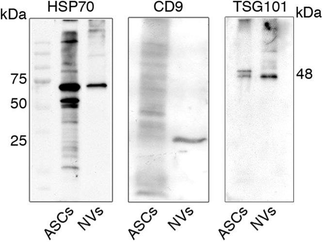
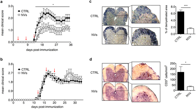
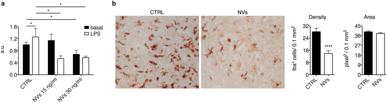
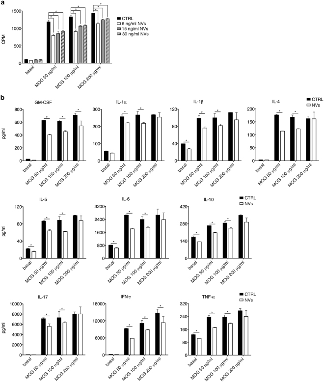
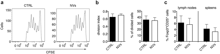
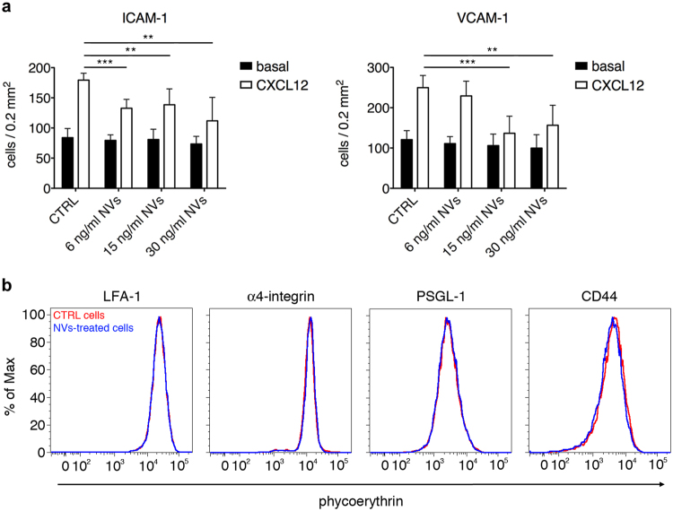
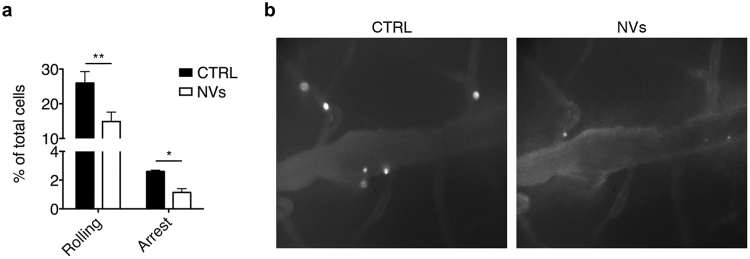
Cell based-therapies represent promising strategies for the treatment of neurological diseases. We have previously shown that adipose stem cells (ASC) ameliorate chronic experimental autoimmune encephalomyelitis (EAE). Recent evidence indicates that most ASC paracrine effects are mediated by extracellular vesicles, i.e. micro- and nanovesicles (MVs and NVs). We show that preventive intravenous administration of NVs isolated from ASC (ASC-NVs) before disease onset significantly reduces the severity of EAE and decreases spinal cord inflammation and demyelination, whereas therapeutic treatment with ASC-NVs does not ameliorate established EAE. This treatment marginally inhibits antigen-specific T cell activation, while reducing microglial activation and demyelination in the spinal cord. Importantly, ASC-NVs inhibited integrin-dependent adhesion of encephalitogenic T cells in vitro, with no effect on adhesion molecule expression. In addition, intravital microscopy showed that encephalitogenic T cells treated with ASC NVs display a significantly reduced rolling and firm adhesion in inflamed spinal cord vessels compared to untreated cells. Our results show that ASC-NVs ameliorate EAE pathogenesis mainly by inhibiting T cell extravasation in the inflamed CNS, suggesting that NVs may represent a novel therapeutic approach in neuro-inflammatory diseases, enabling the safe administration of ASC effector factors.


Conflict of interest statement

The authors declare no competing interests.

Figures

Figure 1
Figure 1
Characterization of ASC-NVs by western blot analysis. Protein lysates from ASC cells and ASC-NV fractions were separated on polyacrylamide gels and probed with specific NVs markers. In NV fractions, a single band at 70 kDa and around to 25 kDa was present after incubation with HSP70 and CD9 antibodies, respectively. Similarly, the treatment with anti-TSG101 antibody displayed an intense signal at 48 kDa in NV populations. Moreover, ASC cells expressed both HSP70 and TSG101 markers whereas showed weak reactivity against CD9 antibody.
Figure 2
Figure 2
Preventive administration of ASC-NVs ameliorates EAE course. (a) MOG35–55-immunized C57Bl/6 mice were treated with PBS (CTRL condition) or 5 μg ASC-NVs at 3, 8 and 13 dpi (red arrows). ASC-NVs significantly inhibited the mean clinical score and chronic EAE development, compared to CTRL condition (day 14 p = 0.041; day 15 p = 0.019; day 16 p = 0.048; day 17 p = 0.045; day 18 p = 0.046; day 19 p = 0.044; day 20 p = 0.033; day 21 p = 0.021; day 22 p = 0.019; day 23 p = 0.020; day 24 p = 0.030; day 25 p = 0.018; day 26 p = 0.018; day 27 p = 0.018; day 28 p = 0.030; day 29 p = 0.049; day 30 p = 0.048; day 31 p = 0.048; day 32 p = 0.041; day 33 p = 0.032; day 34 p = 0.032; day 35 p = 0.024). Data are the mean ± SEM of three independent experiments, for a total of 15 mice/condition. Results are also summarized in Table 1. (b) MOG35–55-immunized C57Bl/6 mice were treated with PBS (CTRL condition) or 5 μg ASC-NVs at 12, 16 and 20 dpi (red arrows). ASC-NVs did not impact EAE development when injected after disease onset. Data are the mean ± SEM of 9 mice/condition. (c) Woelcke staining of lumbar spinal cords showed a reduction of demyelination in mice treated with ASC-NVs, compared to control mice (p = 6.19E-06). Such beneficial effect by ASC-NVs is markedly visible in the blow-up. See also Table 1 for quantification. (d) Immunohistochemistry for CD3+ T lymphocytes on serial sections showed a decreased number of infiltrating CD3+ T cells in EAE lesions from mice treated with ASC-NVs, compared to controls (p = 0.02), as evident in the inserts. See also Table 1 for quantification. For (c) and (d), the data are the mean ± SEM of three independent experiments.
Figure 3
Figure 3
ASC-NVs inhibit microglial cell activation in vitro and in vivo. (a) N9 cells were incubated with LPS 100 ng/ml for 24 h and then treated with 15 ng/ml and 30 ng/ml of ASC-NVs for 48 h. ASC-NVs significantly inhibited microglial cell proliferation in vitro (cntrl basal vs cntrl LPS p = 0.035; cntrl basal vs NVs30 p = 0.039; cntrl LPS vs NVs15 p = 0.020; cntrl LPS vs NVs30 p = 0.012). Data are presented as fluorescence arbitrary units (a.u.) relative to the basal condition and are mean ± SD of a representative experiment performed in triplicate. (b) Evaluation of microglial activation in the spinal cord spinal cord of PBS (CTRL) or NV-treated EAE mice at disease peak. Activated microglial cells were identified by immunohistochemistry, following staining with anti-Iba-1 antibody. Treatment with NVs strongly inhibited microglial activation in EAE mice, as evident by the reduced number of Iba-1+ cells in the spinal cord of NV-treated animals (p = 8.11E-06). Data are the mean ± SEM of three independent experiments.
Figure 4
Figure 4
ASC-NVs partially inhibit MOG35–55-specific CD4+ T cell proliferation and cytokine production in vitro. (a) CD4+ cells isolated from lymph nodes and spleens of 2D2-TCR mice were re-stimulated in vitro for 3 days with increasing concentrations of MOG35–55 peptide, in the presence of irradiated antigen-presenting cells and PBS (CTRL condition) or 30, 15 or 6 ng/ml of ASC-NVs. Cell proliferation was assessed by [3H]-thymidine incorporation and expressed as counts per minute (CPM). ASC-NVs partially reduced antigen-specific T cell proliferation in a dose-dependent manner, when compared with control cells (*p < 0.05). Data are the mean ± SEM of three independent experiments performed in triplicate. (b) Secretion of cytokines (pg/ml) in supernatants by proliferating CD4+ T cells was also significantly affected by ASC-NVs, compared to the control condition (*p < 0.05). Data are the mean ± SD of one representative experiment from a series of two with similar results.
Figure 5
Figure 5
ASC-NVs do not impact CD4+ T cell activation in vivo in EAE mice. 15 × 106-CFSE labeled lymph node and spleen cells from 2D2 mice were injected 8 dpi in EAE recipient mice previously treated with two PBS (CTRL) or ASC-NV injections at 3 and 8 dpi. (a) Representative plots from one control and one NV-treated mouse showing the proliferation of exogenous CD4+CFSE+ T cells detected as CFSE dilution from the original T cell population. (b) Samples were analyzed with FlowJo software to quantitatively assess T cell proliferation in recipient mice. No differences were observed between the proliferation of exogenous CD4+ T cells in control or NV-treated animals. Data are the mean ± SD of five mice/condition. (c) Quantification of Foxp3+CD25+ regulatory T cells (Tregs) in draining lymph nodes and spleens of EAE mice. Lymph nodes and spleens were collected at disease peak from EAE mice treated with PBS (CTRL) or ASC-NVs at day +3, +8 and +13 post-immunization (preventive treatment). Treatment with NVs did not impact the amount of Tregs in both lymph nodes and spleens. Data are shown as % of Foxp3+CD25+ Tregs on the total CD3+CD4+ T cell population and are the mean ± SD of 4 mice/condition.
Figure 6
Figure 6
ASC-NVs inhibit activated T cell integrin-dependent adhesion in vitro. (a) MOG35–55-activated splenocytes from 2D2-TCR were treated for 24 hours with PBS (CTRL condition) or increasing doses of ASC-NVs. Cells were then left spontaneously adhere on purified ICAM-1 or VCAM-1 for 20 min. In some experiments, CXCL12 0.5 μM was added for 5 min to analyze chemokine-induced adhesion. Pre-treatment with NVs significantly inhibited the integrin-dependent adhesion of activated T cells induced by chemokine CXCL12, compared to control condition (ICAM-1: cntrl CXCL12 vs NVs6 CXCL12 p = 0.00011; cntrl CXCL12 vs NVs15 CXCL12 p = 0.0049; cntrl CXCL12 vs NVs30 CXCL12 p = 0.0022. VCAM-1: cntrl CXCL12 vs NVs15 CXCL12 p = 0.00054; cntrl CXCL12 vs NVs30 CXCL12 p = 0.0061). Data are the mean ± SD of three independent experiments performed in triplicate. (b) Adhesion molecules expression on MOG-specific activated T cells after treatment with PBS (CTRL cells) of 30 ng/ml ASC-NVs. Red line: CTRL cells. Blue line: NV-treated cells.
Figure 7
Figure 7
ASC-NVs block activated T cell adhesion in vivo in inflamed spinal cord pial vessels in intravital microscopy experiments. MOG35–55-activated splenocytes from 2D2-TCR were treated for 24 hours with PBS (CTRL condition) or 30 ng/ml NVs. Cells were then fluorescently labeled and injected i.v. in EAE recipient mice at disease peak. (a) Pre-treatment with ASC-NVs strongly reduced activated T cell adhesion in spinal cord venules, compared to control cells (rolling p = 0.0089; arrest p = 0.0325). Data are from five independent experiments, for a total of 28 venules/condition. (b) Representative images of spinal cord pial venules acquired during intravital microscopy experiments. Note the reduced number of adhered NV-treated cells, compared to control cells. Cells are the white spots inside venules.

References

    1. Raine, C. S. The neuropathology of multiple sclerosis. In: Raine, C. S., McFarland, H. & Tourtellotte, W. eds Multiple sclerosis 149–172 (London, UK: Chapman & Hall Med, 1997).
    1. Trapp BD, et al. Axonal transection in the lesions of multiple sclerosis. N. Engl. J. Med. 1998;338:278–285. doi: 10.1056/NEJM199801293380502. - DOI - PubMed
    1. Pluchino S, et al. Injection of adult neurospheres induces recovery in a chronic model of multiple sclerosis. Nature. 2003;422:688–694. doi: 10.1038/nature01552. - DOI - PubMed
    1. Pluchino S, et al. Neurosphere-derived multipotent precursors promote long-lasting neuroprotection by an immunomodulatory mechanism. Nature. 2005;436:266–271. doi: 10.1038/nature03889. - DOI - PubMed
    1. Munoz JR, Stoutenger BR, Robinson AP, Spees JL, Prockop DJ. Human stem/progenitor cells from bone marrow promote neurogenesis of endogenous neural stem cells in the hippocampus of mice. Proc. Natl. Acad. Sci. USA. 2005;102:18171–18176. doi: 10.1073/pnas.0508945102. - DOI - PMC - PubMed

Publication types

MeSH terms