

doi: 10.1038/s41375-018-0134-9.
Epub 2018 Apr 19.
GATA2 monoallelic expression underlies reduced penetrance in inherited GATA2-mutated MDS/AML
Affiliations
- PMID: 29749400
- PMCID: PMC6224398
- DOI: 10.1038/s41375-018-0134-9
Item in Clipboard
GATA2 monoallelic expression underlies reduced penetrance in inherited GATA2-mutated MDS/AML
Leukemia.
2018 Nov.
No abstract available
Conflict of interest statement
The authors declare that they have no conflict of interest.
Figures
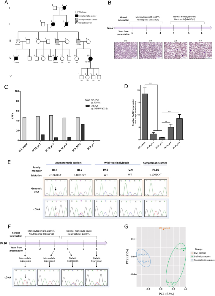
Investigating the molecular mechanisms underlying the reduced penetrance of germline p.Thr354Met mutations observed in a GATA2-mutated MDS/AML family. a Genogram of the GATA2-mutated pedigree. Squares denote males and circles denote females. This five-generation MDS/AML family presented to Barts Health hospital in London with identical germline GATA2 mutations (p.Thr354Met; c.1061C>T) and variable clinical manifestations. Two first-degree cousins (IV.1 and IV.6) presented at 23 and 18 years of age, respectively, with high-grade MDS transforming to AML and monosomy 7. Both cousins died post allogeneic hematopoietic stem cell transplant (HSCT) due to transplant-related complications (IV.1 from graft vs. host disease (GvHD) and IV.6 from relapsed MDS/AML). Ten years later, their first cousin (IV.10) developed symptoms at 31 years, including recurrent minor infections and significant leukopenia (monocytopenia [0.1 × 10⁹/L] and neutropenia [0.8 × 10⁹/L]) with mild macrocytosis and normal hemoglobin and platelet counts. She remains under close surveillance where her blood counts are routinely monitored. All four of her children have inherited her WT GATA2 allele. Similarly, members (IV.7, IV.8, and IV.9) were screened for the mutation and all have a WT GATA2 configuration. The paternal grandmother (II.2) of IV.10 as well as her paternal great-uncle (II.3) and great-grandmother (I.2) all were reported to have died of AML (ages of disease onset were 53, 24, and 53-years old, respectively). Not only did GATA2 mutations correlate with early age of disease onset in the fourth generation (IV.1/23 yr., IV.6/18 yr., and IV.10/31 yr.), but the parental third-generation carriers (III.1, III.5, and III.7) remain hematologically normal and symptom-free into their mid–late 60s. No material was available from other family members. b A clinical timeline of IV.10 showing the change in clinical parameters over the course of disease presentation. Photographs of peripheral blood smears from IV.10 (yr. 1, 3, 4, and 6) stained with May-Grünwald Giemsa staining. Magnification: ×20. c Secondary ASXL1 mutations: variant allele frequencies of GATA2 germline mutation and ASXL1 acquired mutation. Samples from three individuals were sequenced: one asymptomatic parent (III.7), one deceased MDS/AML cousin (IV.6), and across three time-points (yr. 1, 4, and 6) from the symptomatic patient (IV.10) reflecting disease evolution. d
GATA2 global expression measured by qRT-PCR of bone marrow samples and normalized to healthy bone marrow control: downregulation in IV.10_yr.1 compared with III.7 and downregulation in IV.10_yr.1–3 GATA2 expression compared with IV.10_yr.4–6. The average of five independent experiments is shown. Statistical significance was determined at *p < 0.05, **p < 0.01, and ***p < 0.001 using a t-test with Bonferroni correction. Error bars represent standard error of the mean (SEM). e
GATA2 monoallelic expression of the mutant allele in symptomatic (IV.10) vs. asymptomatic carriers (III.5 and III.7), as measured by cDNA sequencing of bone marrow samples. f Correlation of monoallelic GATA2 expression with disease symptoms across the time-points studied in IV.10 with reactivation of the WT allele “C” expression noted 3 years after presentation, concurrent with improvements in hematological parameters. g RNA-seq analysis: principal component analysis (PCA) plot showing a good separation between GATA2 biallelic (green) and monoallelic (blue) groups based on all transcriptomes
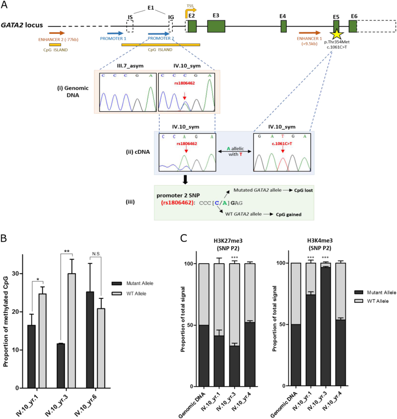
Elucidating the molecular mechanisms driving allele-specific changes in GATA2 expression. a(i) A noncoding SNP (rs1806462 [C/A]) located within the second GATA2 promoter region overlapping a CpG island was detected in the symptomatic (IV.10) but not in asymptomatic members (III.7). a(ii) Given the location of promoter 2 SNP within the 5’UTR, a haplotype between the SNP allele “A” and the germline mutant allele “T” was established, providing a means of distinguishing between mutant and WT alleles in subsequent experiments. a(iii) This promoter SNP [C/A] removes a CpG methylation site in the mutant allele “A” and generates a CpG methylation site in the WT allele “C”. b The proportion of methylated CpGs between mutant and WT alleles across the three time-points of IV.10. WT allele is significantly more methylated than the mutant allele in monoallelic samples (yr. 1 and yr. 3), whereas no significant allele-specific differences in methylation were observed in a biallelic-expressing sample (yr. 6). The average of three independent experiments is shown. c Quantification of mutant and WT allele ChIP sequence peak heights across the time-points of IV.10 based on Sanger sequencing. H3K4me3 activation mark favoring the mutant allele was enriched in monoallelic samples (yr. 1 and yr. 3) compared with the biallelic sample (yr. 4). The average of three independent experiments is shown. Statistical significance was determined at *p < 0.05, **p < 0.01, and ***p < 0.001 using a t-test with Bonferroni correction. NS corresponds to nonsignificant comparisons. Error bars represent SEM
References
Publication types
MeSH terms
Substances
Grants and funding
LinkOut - more resources
Full Text Sources
Other Literature Sources
Medical
Molecular Biology Databases
Research Materials
Miscellaneous
