Review
. 2018 Aug;42(2):691-702.
doi: 10.3892/ijmm.2018.3668. Epub 2018 May 10.

Personalized regulation of glioblastoma cancer stem cells based on biomedical technologies: From theory to experiment (Review)

Affiliations
Review

Personalized regulation of glioblastoma cancer stem cells based on biomedical technologies: From theory to experiment (Review)

Igor Bryukhovetskiy et al. Int J Mol Med. 2018 Aug.

Abstract

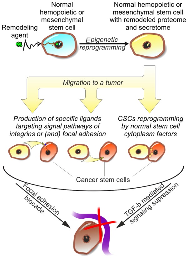
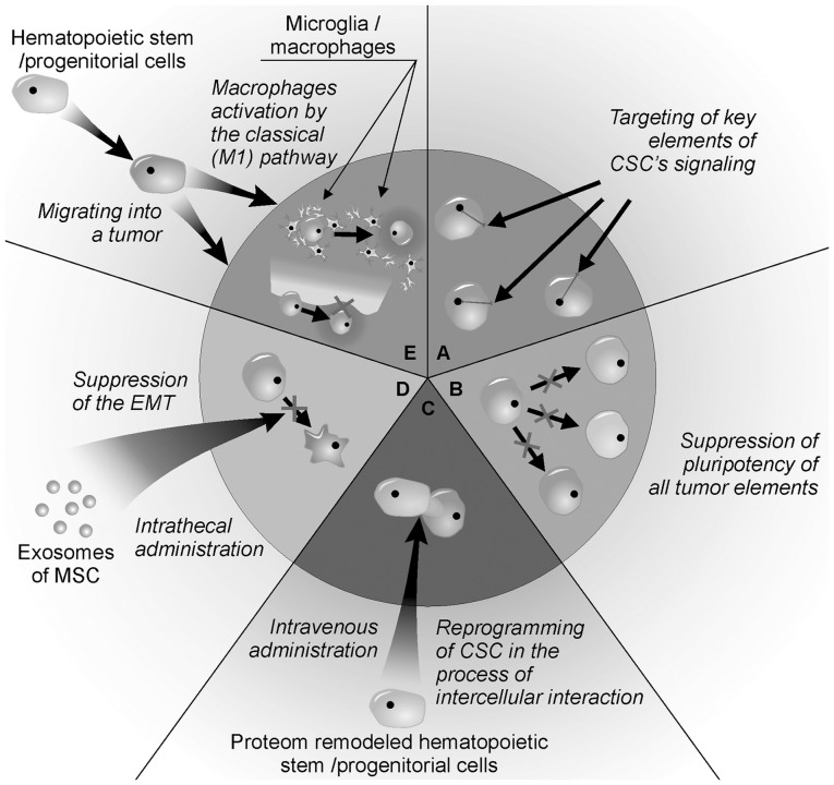
Glioblastoma multiforme (GBM) is one of the most aggressive brain tumors. GBM represents >50% of primary tumors of the nervous system and ~20% of intracranial neoplasms. Standard treatment involves surgery, radiation and chemotherapy. However, the prognosis of GBM is usually poor, with a median survival of 15 months. Resistance of GBM to treatment can be explained by the presence of cancer stem cells (CSCs) among the GBM cell population. At present, there are no effective therapeutic strategies for the elimination of CSCs. The present review examined the nature of human GBM therapeutic resistance and attempted to systematize and put forward novel approaches for a personalized therapy of GBM that not only destroys tumor tissue, but also regulates cellular signaling and the morphogenetic properties of CSCs. The CSCs are considered to be an informationally accessible living system, and the CSC proteome should be used as a target for therapy directed at suppressing clonal selection mechanisms and CSC generation, destroying CSC hierarchy, and disrupting the interaction of CSCs with their microenvironment and extracellular matrix. These objectives can be achieved through the use of biomedical cellular products.


Figures

Figure 1
Figure 1
Electron microscopy examination of human glioblastoma U87MG cells, indicating the mechanisms of glioblastoma cell interaction, examined by the authors. (A) Fusion of two interacting cells (magnification, ×2,300). (B) Numerous mergers between cells (magnification, ×953). (C) Conglomerate forming from interacting cells (magnification, ×793). (D) Creation of close contacts among the cells with interdigitations (magnification, ×13,380). (E) formation of gap junctions (magnification, ×40,150). (F) subsequent dissolution of cytomembrane (magnification, ×28,600); (G) Special differentiation of cytomembrane into microtubes and/or connective bridges (magnification, ×493). (H) Formation of microtubes between remote cells (magnification, ×919). (I) Microtubes formation between close cells (magnification, ×798).
Figure 2
Figure 2
Fluorescent laser microscopy examination of glioblastoma U87MG cell culture, examined by the authors. Green tag (arrows labeled 1) represents CD133+ cells stained with Vybrant® CFDA SE cell tracer (V12883; Molecular Probes; Thermo Fisher Scientific, Inc., Waltham, MA, USA; λ=488 nm; 25 µM in PBS for 25 min at 37°С). Red tag (arrows labeled 2) represents СD44+ cells stained with CellTracker™ Red CMTPX Dye (C34552; Molecular Probes; Thermo Fisher Scientific, Inc.; λ=546 nm; 15 µM in DMEM for 25 min at 37°С). Staining was conducted according to the manufacturer's protocol. After 48 h, CD133+ showed red fluorescent tag indicating the presence of cytoplasm components, CD44+ cells (arrows labeled 3).
Figure 3
Figure 3
Fluorescent laser microscopy examination of combined culture of stem and cancer cells of human glioblastoma, examined by the authors. Green tag represents hematopoietic stem cells stained with Vybrant® CFDA SE tracer (V12883; Molecular Probes; Thermo Fisher Scientific, Inc.; λ=488 nm; 25 µM in PBS for 25 min at 37°С). Red tag represents glioblastoma cells stained with CellTracker™ Red CMTPX Dye (C34552; Molecular Probes; Thermo Fisher Scientific, Inc.; λ=546 nm; 15 µM in DMEM for 25 min at 37°С). (А) Adhesion of HSCs to cancer cells; (B) fluorescent tag exchange; components of fluorescent tag (C) on the surface and (D) in the cytoplasm of cancer cells.
Figure 4
Figure 4
Scheme of personalized therapy for glioblastoma multiforme involving hematopoietic stem cells with remodeled proteome and secretome. Cellular therapy results in breaking the connection between CSCs and extracellular matrix, impeding invasion. CSCs, cancer stem cells; TGF-β, transforming growth factor β.
Figure 5
Figure 5
Methods of personalized regulation of glioblastoma cancer stem cells. (A) Identification of molecular targets in CSC proteome and their inhibition with chemotherapeutic agents. (B) Suppression of pluripotency in all types of CSCs via using inhibitors of Wnt-dependent signaling pathway. (C) Reprogramming of CSCs during their interaction with normal stem cells. (D) Inhibition of EMT in CSCs using exosomes of MSCs. (E) Destabilization of CSC interaction with local microsurroundings and extracellular matrix via monocytes and macrophages with a set vector of classic (pro-inflammatory) pathway activation. CSC, cancer stem cell; EMT, epithelial-mesenchymal transition; MSC, mesenchymal stem cell.

References

    1. Stupp R, Toms SA, Kesari S. Treatment for patients with newly diagnosed glioblastoma-reply. JAMA. 2016;315:2348–2349. doi: 10.1001/jama.2016.1847. - DOI - PubMed
    1. Omuro A, DeAngelis LM. Glioblastoma and other malignant gliomas: A clinical review. JAMA. 2013;310:1842–1850. doi: 10.1001/jama.2013.280319. - DOI - PubMed
    1. Louis DN, Perry A, Reifenberger G, von Deimling A, Figarella-Branger D, Cavenee WK, Ohgaki H, Wiestler OD, Kleihues P, Ellison DW. The 2016 World Health Organization classification of tumors of the central nervous system: A summary. Acta Neuropathol. 2016;131:803–820. doi: 10.1007/s00401-016-1545-1. - DOI - PubMed
    1. DeWitt JC, Mock A, Louis DN. The 2016 WHO classification of central nervous system tumors: What neurologists need to know. Curr Opin Neurol. 2017;30:643–649. doi: 10.1097/WCO.0000000000000490. - DOI - PubMed
    1. Louis DN, Perry A, Burger P, Ellison DW, Reifenberger G, von Deimling A, Aldape K, Brat D, Collins VP, Eberhart C, et al. International society of Neuropathology-haarlem consensus guidelines for nervous system tumor classification and grading. Brain Pathol. 2014;24:429–435. doi: 10.1111/bpa.12171. - DOI - PMC - PubMed