

Contemporary understanding of the secretory granules in human eosinophils
- PMID: 29749658
- PMCID: PMC6013358
- DOI: 10.1002/JLB.3MR1217-476R
Contemporary understanding of the secretory granules in human eosinophils
Abstract
Eosinophil secretory (specific) granules have a unique morphology and are both a morphologic hallmark of eosinophils and fundamental to eosinophil-mediated responses. Eosinophil mediators with multiple functional activities are presynthesized and stored within these granules, poised for very rapid, stimulus-induced secretion. The structural organization and changes of eosinophil specific granules are revealing in demonstrating the complex and diverse secretory activities of this cell. Here, we review our current knowledge on the architecture, composition, and function of eosinophil specific granules as highly elaborated organelles able to produce vesiculotubular carriers and to interplay with the intracellular vesicular trafficking. We reconsider prior identifications of eosinophil cytoplasmic granules, including "primary," "secondary," "microgranules," and "small granules"; and consonant with advances, we provide a contemporary recognition that human eosinophils contain a single population of specific granules and their developmental precursors and derived secretory vesicles.
Keywords: Eosinophil Sombrero Vesicles; cell secretion; degranulation; immune responses; inflammation; transmission electron microscopy; vesicular trafficking.
©2018 Society for Leukocyte Biology.
Conflict of interest statement
The authors declare no conflict of interest.
Figures
Similar articles
-
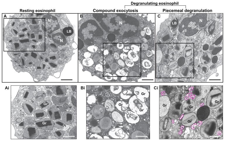
CD63 is tightly associated with intracellular, secretory events chaperoning piecemeal degranulation and compound exocytosis in human eosinophils.J Leukoc Biol. 2016 Aug;100(2):391-401. doi: 10.1189/jlb.3A1015-480R. Epub 2016 Mar 10. J Leukoc Biol. 2016. PMID: 26965633 Free PMC article.
-
Vesicle-mediated secretion of human eosinophil granule-derived major basic protein.Lab Invest. 2009 Jul;89(7):769-81. doi: 10.1038/labinvest.2009.40. Epub 2009 Apr 27. Lab Invest. 2009. PMID: 19398958 Free PMC article.
-
Vesicular trafficking of immune mediators in human eosinophils revealed by immunoelectron microscopy.Exp Cell Res. 2016 Oct 1;347(2):385-90. doi: 10.1016/j.yexcr.2016.08.016. Epub 2016 Aug 22. Exp Cell Res. 2016. PMID: 27562864 Free PMC article. Review.
-
Mechanisms of eosinophil secretion: large vesiculotubular carriers mediate transport and release of granule-derived cytokines and other proteins.J Leukoc Biol. 2008 Feb;83(2):229-36. doi: 10.1189/jlb.0707503. Epub 2007 Sep 17. J Leukoc Biol. 2008. PMID: 17875811 Free PMC article. Review.
-
Expression and subcellular localization of the Qa-SNARE syntaxin17 in human eosinophils.Exp Cell Res. 2015 Oct 1;337(2):129-135. doi: 10.1016/j.yexcr.2015.07.003. Epub 2015 Aug 6. Exp Cell Res. 2015. PMID: 26254897 Free PMC article.
Cited by
-
Eosinophils promote pulmonary matrix destruction and emphysema via Cathepsin L.Signal Transduct Target Ther. 2023 Oct 11;8(1):390. doi: 10.1038/s41392-023-01634-x. Signal Transduct Target Ther. 2023. PMID: 37816708 Free PMC article.
-
Eosinophil ETosis-Mediated Release of Galectin-10 in Eosinophilic Granulomatosis With Polyangiitis.Arthritis Rheumatol. 2021 Sep;73(9):1683-1693. doi: 10.1002/art.41727. Epub 2021 Aug 11. Arthritis Rheumatol. 2021. PMID: 33750029 Free PMC article.
-
Systems Biology Methods via Genome-Wide RNA Sequences to Investigate Pathogenic Mechanisms for Identifying Biomarkers and Constructing a DNN-Based Drug-Target Interaction Model to Predict Potential Molecular Drugs for Treating Atopic Dermatitis.Int J Mol Sci. 2024 Oct 4;25(19):10691. doi: 10.3390/ijms251910691. Int J Mol Sci. 2024. PMID: 39409019 Free PMC article.
-
Eosinophil extracellular vesicles and DNA traps in allergic inflammation.Front Allergy. 2024 Aug 1;5:1448007. doi: 10.3389/falgy.2024.1448007. eCollection 2024. Front Allergy. 2024. PMID: 39148911 Free PMC article. Review.
-
Charcot-Leyden Crystals in Eosinophilic Inflammation: Active Cytolysis Leads to Crystal Formation.Curr Allergy Asthma Rep. 2019 Jun 15;19(8):35. doi: 10.1007/s11882-019-0868-0. Curr Allergy Asthma Rep. 2019. PMID: 31203469 Free PMC article. Review.
References
-
- Stow JL, Murray RZ. Intracellular trafficking and secretion of inflammatory cytokines. Cytokine Growth Factor Rev. 2013;24:227–39. - PubMed
-
- Melo RCN, Dvorak AM, Weller PF. Eosinophil Ultrastructure. In: Lee J, Rosenberg H, editors. Eosinophils in health and disease. Vol. 1. Elsevier; New York: 2012. pp. 20–27.
Publication types
MeSH terms
Grants and funding
LinkOut - more resources
Full Text Sources
Other Literature Sources
Molecular Biology Databases
