

The Role of Neuroimaging in the Determination of Brain Death
- PMID: 29749664
- PMCID: PMC6055878
- DOI: 10.1111/jon.12516
The Role of Neuroimaging in the Determination of Brain Death
Abstract
Background and purpose: Brain death determination (BDD) is primarily a clinical diagnosis, where death is defined as the permanent loss of brainstem function. In scenarios where clinical examinations are inaccurate, ancillary imaging tests are required. The choice of ancillary imaging test is variable, but the common denominator for all of them is to establish a lack of cerebral blood flow. The purpose of this study was to compare the diagnostic accuracy and interrater reliability of different ancillary imaging tests used for BDD.
Methods: Archival data were retrospectively analyzed for all patients who underwent any ancillary imaging test for BDD at our institution. The results of ancillary imaging tests were compared with, the reference standard, the clinical checklist for declaration of brain death. Sensitivity, specificity, positive predictive value (PPV), and negative predictive value (NPV) of different ancillary imaging tests for BDD were performed. Interobserver agreement between two observers was measured using kappa statistics for each of the imaging modalities.
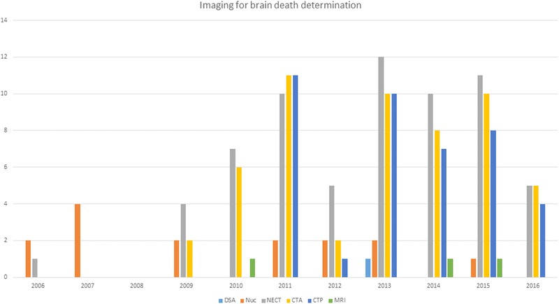
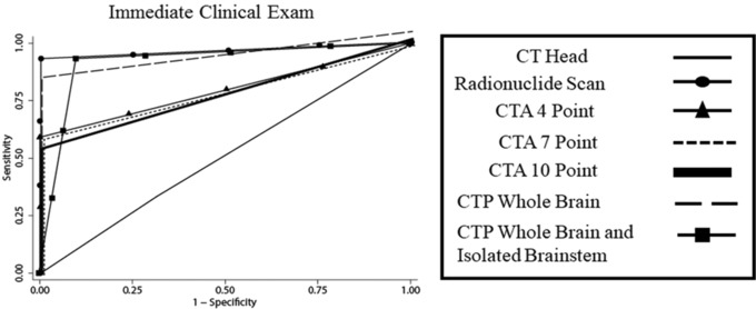
Results: A total of 74 patients underwent 41 computer tomography perfusion (CTP), 54 CT angiogram, 15 radionuclide scans, 1 cerebral angiogram, 3 magnetic resonance imaging, and 71 nonenhanced CT (NECT) head for BDD. All ancillary tests (except NECT head) showed 100% specificity and PPV. CTP had the highest sensitivity and NPV. All ancillary imaging tests demonstrated very high interrater reliability.
Conclusions: The uses of ancillary imaging tests for BDD are increasing. Within this study's limitations, CTP followed by radionuclide scan were found to be the most accurate and reliable ancillary imaging test for BDD.
Keywords: CT perfusion; Imaging; brain death.
© 2018 The Authors. Journal of Neuroimaging published by Wiley Periodicals, Inc. on behalf of American Society of Neuroimaging.
Figures
References
-
- Shemie SD, Lee D, Sharpe M, et al. Brain blood flow in the neurological determination of death: Canadian expert report. Can J Neurol Sci 2008;35:140‐5. - PubMed
-
- Wijdicks EFM. The diagnosis of brain death. N Engl J Med 2001;344:1215‐21. - PubMed
-
- Wijdicks EFM. Brain death worldwide: accepted fact but no global consensus in diagnostic criteria. Neurology 2002;58:20‐5. - PubMed
MeSH terms
LinkOut - more resources
Full Text Sources
Other Literature Sources
