

The Preventive Role of Pioglitazone in Glycerol-Induced Acute Kidney Injury in Rats during Two Different Treatment Periods
- PMID: 29749987
- PMCID: PMC5936850
The Preventive Role of Pioglitazone in Glycerol-Induced Acute Kidney Injury in Rats during Two Different Treatment Periods
Abstract
Background: Acute kidney injury is the most life-threatening complication of rhabdomyolysis. Glycerol is commonly used to induce this injury. The aim was to investigate the renoprotective effects of pioglitazone and the possible advantage of administering the drug for a longer period.
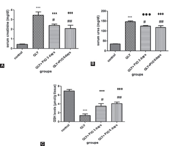
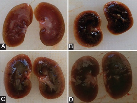
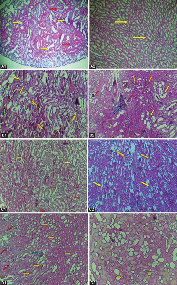
Methods: Twenty-four male Albino Wistar rats were randomly divided into 4 groups (n=6/group): (A) control, (B) glycerol (50%, 10 mL/kg intramuscularly), (C) glycerol+pioglitazone (10 mg/kg orally for 3 days), and (D) glycerol+pioglitazone (for 6 days). Serum urea and creatinine levels were measured to assess the renal function. Reduced glutathione (GSH) levels and histological alterations were also measured. Statistical analysis was performed using Prism (version 6). The numerical data were evaluated by ANOVA, followed by the Tukey tests. The categorical data were evaluated by the Mann-Whitney test and the Fisher exact tests. P<0.05 was considered significant.
Results: In the glycerol-injected rats, the serum urea and creatinine levels were increased (P<0.001), while the GSH levels were decreased (P<0.001) compared to Group A. The nephrotoxicity showed significant tubular (P=0.01) and glomerular (P=0.02) injuries. In the pioglitazone-treated rats, the changes in the serum biomarkers and in the GSH levels were reversed in Group C (P=0.01) and in Group D (P=0.01). The microscopic examinations of the kidneys also showed some improvement. No obvious statistically significant difference was found between these 2 preventive groups in most studied features.
Conclusion: These results indicate that pioglitazone might have nephroprotective effects in this injury model. Pioglitazone succeeded in producing this effect within 3 days. Doubling the drug administration period did not produce any significant superior benefit.
Keywords: Acute kidney injury; Glycerol; Kidney; Pioglitazone; Reduced glutathione; Rhabdomyolysis.
Figures
References
-
- Petejova N, Martinek A. Acute kidney injury due to rhabdomyolysis and renal replacement therapy: a critical review. Crit Care. 2014;18:224. doi: 10.1186/cc13897. [ PMC Free Article] - DOI - PMC - PubMed
-
- Torres PA, Helmstetter JA, Kaye AM, Kaye AD. Rhabdomyolysis: pathogenesis, diagnosis, and treatment. Ochsner J. 2015;15:58–69. [ PMC Free Article] - PMC - PubMed
LinkOut - more resources
Full Text Sources