. 2017 Jul;1(7):0084.
doi: 10.1038/s41551-017-0084. Epub 2017 Jun 26.

Light-sheet microscopy for slide-free non-destructive pathology of large clinical specimens

Affiliations

Light-sheet microscopy for slide-free non-destructive pathology of large clinical specimens

Adam K Glaser et al. Nat Biomed Eng. 2017 Jul.

Abstract

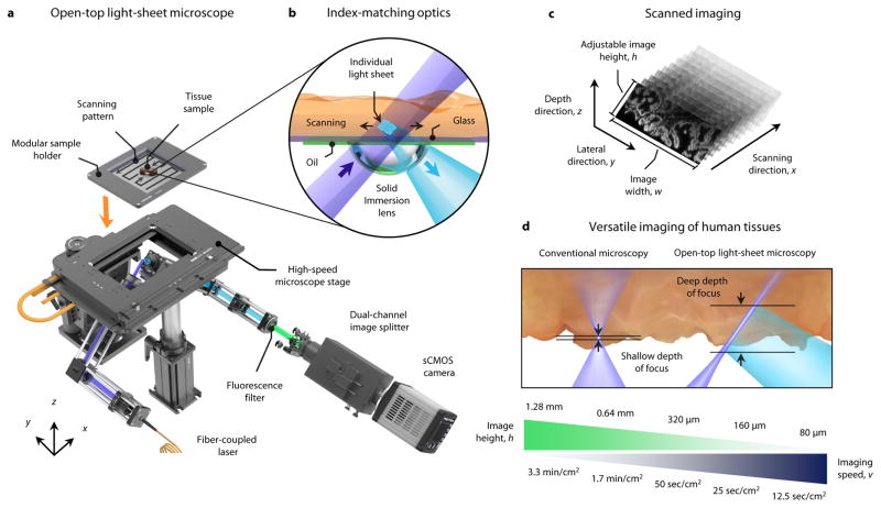
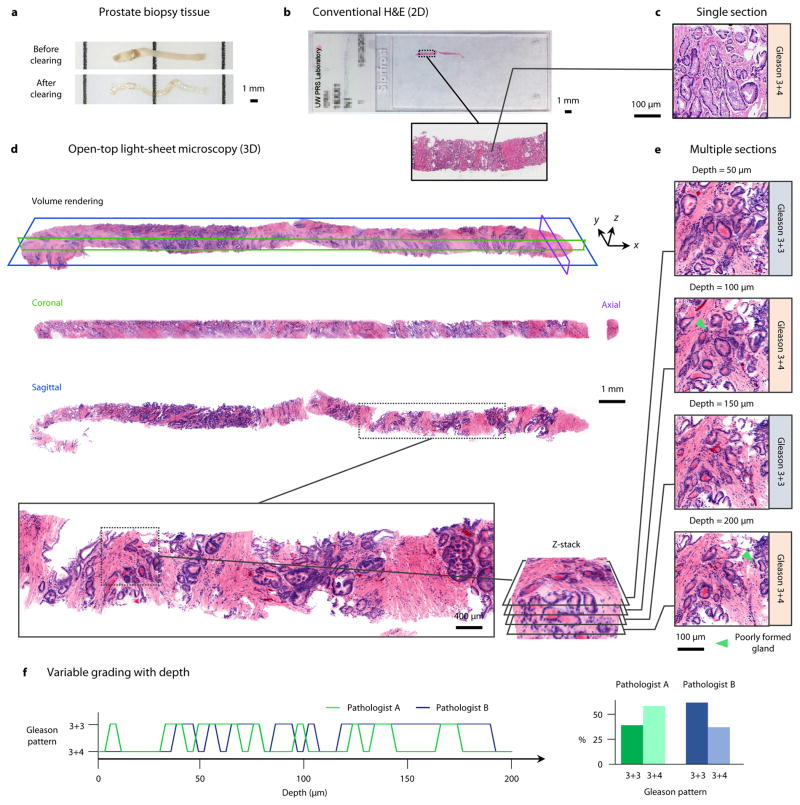
For the 1.7 million patients per year in the U.S. who receive a new cancer diagnosis, treatment decisions are largely made after a histopathology exam. Unfortunately, the gold standard of slide-based microscopic pathology suffers from high inter-observer variability and limited prognostic value due to sampling limitations and the inability to visualize tissue structures and molecular targets in their native 3D context. Here, we show that an open-top light-sheet microscope optimized for non-destructive slide-free pathology of clinical specimens enables the rapid imaging of intact tissues at high resolution over large 2D and 3D fields of view, with the same level of detail as traditional pathology. We demonstrate the utility of this technology for various applications: wide-area surface microscopy to triage surgical specimens (with ~200 μm surface irregularities), rapid intraoperative assessment of tumour-margin surfaces (12.5 sec/cm2), and volumetric assessment of optically cleared core-needle biopsies (1 mm in diameter, 2 cm in length). Light-sheet microscopy can be a versatile tool for both rapid surface microscopy and deep volumetric microscopy of human specimens.


Conflict of interest statement

Competing interests The authors declare no competing financial interests.

Figures

Figure 1
Figure 1. Open-top light-sheet microscope for clinical pathology
a, An illumination light sheet enters the bottom surface of a tissue sample at an oblique 45-deg angle (purple). The specimen(s) is placed on a modular glass-plate sample holder, which is inserted into a two-axis translation stage and scanned in a serpentine pattern of volumetric image strips to enable 3D imaging over a large lateral extent. Fluorescence emission (cyan), which is generated along the light sheet, is collected in the orthogonal direction by an objective lens. The fluorescence signal is then transmitted through an emission filter (green) and a dual-channel image splitter (for 2 color imaging) before being imaged onto a high-speed sCMOS camera. b, To provide aberration-free imaging, a solid immersion lens (SIL) and oil layer are used for refractive-index matching of both the illumination and collection beams into and out of the glass plate and tissue sample. c, As the sample is translated in the primary scanning direction, x, oblique 2D light-sheet images with a width, w, and adjustable height, h, are captured in succession to form a 3D imaging volume. d, In contrast to conventional microscopy methods that have a shallow fixed depth of focus and slow 3D imaging rates, the deep depth of focus and adjustable vertical field of view of the open-top light-sheet microscope makes it optimal for both rapid microscopy of irregular/tilted tissue surfaces, and deep volumetric microscopy of clinical specimens. The imaging speeds shown correspond to acquiring single-channel images with height, h.
Figure 2
Figure 2. Wide-area surface microscopy of fresh human prostate
a, In preparation for post-operative pathological evaluation, radical prostatectomy specimens are grossly processed by manually cutting the organ into cross-sectional slices (3 to 5 mm in thick). b, A photograph of a fresh prostate slice (3.1 × 3.5 × 0.4 cm) imaged with the open-top light-sheet microscope. c, The tissues are stained with acridine orange for 20 sec and then imaged at a speed of v = 50 sec/cm2, which provides a vertical field of view of h = 320 μm to accommodate for any surface irregularities and tilting errors. A horizontal (en face) 2D “section” from the 3D dataset is shown on the top left, prior to surface extraction, where regions of defocus and incomplete imaging are seen (inset arrows). On the bottom right, the irregular surface of the large specimen has been digitally extracted from the 3D dataset to provide a comprehensive image of the surface. d, A depth profile of the tissue surface, along the dashed pink line, reveals significant surface irregularities (up to 200 μm) as well as a slight tilt in the glass-plate sample holder (2 μm/mm or 0.2% slope). e, A histogram of the tissue surface depth from all N=25 prostate slices imaged in this study (average and 95% confidence intervals, CI). For illustrative purposes, the depth of focus is indicated for both a conventional single-axis microscope and our open-top light-sheet microscope. f, Moderate- and high-magnification images of normal prostate glands, where a layer of both basal and epithelial cells is observed (inset arrows). A corresponding H&E histology image is shown on the right. g, A region with benign prostate (left) transitioning into prostate adenocarcinoma (right), which exhibits a crowding of glands with a single epithelial cell layer (inset arrow). h, High-grade prostate intraepithelial neoplasia, displaying stratified nuclei and tufted projections into the lumen. A corresponding H&E histology image is shown on the right.
Figure 3
Figure 3. Rapid intra-operative microscopy of fresh human breast
a,b, A freshly excised specimen of human breast tissue (2.0 × 2.0 × 0.4 cm) was stained with acridine orange for 20 sec and then imaged with the open-top light-sheet microscope at a speed of v = 12.5 sec/cm2. The tissue was subsequently submitted for conventional histology. c, A surface-extracted image acquired in <1 min with representative line profiles of the tissue-surface depth, d. In (a–c) the inset arrows denote a region of invasive carcinoma (i.e. a positive margin). e, Moderate- and high-magnification images reveal a transition from benign breast tissue to invasive ductal carcinoma. f, Benign breast lobules are clearly visualized (inset arrows) and correlate with conventional histology. g, Open-top light-sheet microscopy, conventional histology, and frozen-sectioning of fibro-adipose tissue. Open-top light-sheet microscopy images and FFPE histology sections both reveal adipocytes and strands of fibrous tissue with stromal cells, whereas the frozen section contains distorted fibrous tissue and adipocytes that are distorted by the process (inset arrows).
Figure 4
Figure 4. Volumetric dual-channel imaging of a cleared human biopsy
a, A 2-cm long by 1-mm diameter human prostate core-needle biopsy, before and after optical clearing (overnight procedure). b, A conventional H&E-stained slide of a single 5-μm thick section of the biopsy sample. c, A high-magnification view of a Gleason 3+4 carcinoma region from the histology slide. d, The prostate biopsy was stained with DRAQ5 and eosin, and false-colored to mimic the traditional H&E color palette. A volume rendering of the biopsy is shown, which can be digitally “sectioned” into orthogonal 2D cross sections in the coronal (green), axial (purple), and sagittal (blue) planes. e, A high-magnification view of one sagittal region of interest, visualized at multiple depths (5 μm increments) to a depth of 200 μm. Gleason grading is found to differ between 3+3 and 3+4 due to tangential sectioning artifacts creating the appearance of poorly formed glands (inset arrows). f, Grading of the region of interest as a function of depth (5-μm increments) by two pathologists. When assessing individual depths, the grading varies between 3+3 (40% for Pathologist A, 62.5% for Pathologist B) and 3+4 (60% for Pathologist A, 37.5% for Pathologist B), with inter-observer agreement of 47.5%. However, after comprehensive review of the entire volumetric dataset, both pathologists agreed upon an unambiguous diagnosis of Gleason 3+3 based on the observation that structures which appeared to be “poorly formed” pattern 4 glands were tangential-sectioning artifacts of well-formed pattern 3 glands.

References

    1. Surveillance Research Program, N.C.I. [12-13-2016];Fast Stats: An interactive tool for access to SEER cancer statistics. 2016 Available from: https://seer.cancer.gov/faststats/
    1. Barakat FH, Sulaiman I, Sughayer MA. Reliability of frozen section in breast sentinel lymph node examination. Breast Cancer. 2014;21(5):576–82. - PubMed
    1. McKenney JK, et al. The potential impact of reproducibility of Gleason grading in men with early stage prostate cancer managed by active surveillance: a multi-institutional study. J Urol. 2011;186(2):465–9. - PubMed
    1. Shah RB, et al. Diagnosis of Gleason pattern 5 prostate adenocarcinoma on core needle biopsy: an interobserver reproducibility study among urologic pathologists. Am J Surg Pathol. 2015;39(9):1242–9. - PubMed
    1. Meyer JS, et al. Breast carcinoma malignancy grading by Bloom-Richardson system vs proliferation index: reproducibility of grade and advantages of proliferation index. Mod Pathol. 2005;18(8):1067–78. - PubMed