. 2018 May 9;4(5):eaao5838.
doi: 10.1126/sciadv.aao5838. eCollection 2018 May.

Mammalian EAK-7 activates alternative mTOR signaling to regulate cell proliferation and migration

Affiliations

Mammalian EAK-7 activates alternative mTOR signaling to regulate cell proliferation and migration

Joe Truong Nguyen et al. Sci Adv. .

Abstract

Nematode EAK-7 (enhancer-of-akt-1-7) regulates dauer formation and controls life span; however, the function of the human ortholog mammalian EAK-7 (mEAK-7) is unknown. We report that mEAK-7 activates an alternative mechanistic/mammalian target of rapamycin (mTOR) signaling pathway in human cells, in which mEAK-7 interacts with mTOR at the lysosome to facilitate S6K2 activation and 4E-BP1 repression. Despite interacting with mTOR and mammalian lethal with SEC13 protein 8 (mLST8), mEAK-7 does not interact with other mTOR complex 1 (mTORC1) or mTOR complex 2 (mTORC2) components; however, it is essential for mTOR signaling at the lysosome. This phenomenon is distinguished by S6 and 4E-BP1 activity in response to nutrient stimulation. Conventional S6K1 phosphorylation is uncoupled from S6 phosphorylation in response to mEAK-7 knockdown. mEAK-7 recruits mTOR to the lysosome, a crucial compartment for mTOR activation. Loss of mEAK-7 results in a marked decrease in lysosomal localization of mTOR, whereas overexpression of mEAK-7 results in enhanced lysosomal localization of mTOR. Deletion of the carboxyl terminus of mEAK-7 significantly decreases mTOR interaction. mEAK-7 knockdown decreases cell proliferation and migration, whereas overexpression of mEAK-7 enhances these cellular effects. Constitutively activated S6K rescues mTOR signaling in mEAK-7-knocked down cells. Thus, mEAK-7 activates an alternative mTOR signaling pathway through S6K2 and 4E-BP1 to regulate cell proliferation and migration.


Figures
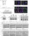
Fig. 1
Fig. 1. mEAK-7 is a lysosomal protein, conserved across eukaryotes, and is required for mTOR signaling in human cells.
(A) Comparison of eukaryotic mEAK-7 orthologs. (B) Diagram depicting the mEAK-7 N-myristoylation motif and the TLD (TBC/LysM-associated) domain. (C) Immunoblot screen of human cell lines to detect mEAK-7 protein. (D) Confocal microscopy analysis of H1299 cells stably expressing HA–mEAK-7WT for HA and LAMP2 or LAMP1. Scale bars, 10 μm. (E) Analysis of endogenous colocalization of mEAK-7 and lysosomal markers in H1299 cells. Scale bars, 25 μm. (F) H1975 cells were treated with control (Ctl) or three unique mEAK-7 siRNAs to assess S6 phosphorylation. (G) H1975, MDA-MB-231, and H1299 cells were treated with control (Ctl) or two unique mEAK-7 siRNAs to assess S6 phosphorylation. (H) Cells were treated with control (Ctl) or mEAK-7 #1 siRNA to assess 4E-BP1 phosphorylation. (I) Cells were treated with control or two unique mEAK-7 siRNAs. Next, cells were starved in DMEM−AAs for 2 hours, and amino acids, insulin, or both were reintroduced for 30 min. (J) Cells were treated with control, mEAK-7 #1, S6K1, and S6K2 siRNA. Next, cells were starved in DMEM+AAs for 2 hours, and insulin (1 and 10 μM) were reintroduced for 30 min. All experiments were repeated at least three times. Glyceraldehyde-3-phosphate dehydrogenase (GAPDH) was used for loading controls. hESC, human embryonic stem cells.
Fig. 2
Fig. 2. Overexpression of mEAK-7 activates mTOR signaling and the TBC/LysM-associated domain and mTOR-binding domain are necessary for mEAK-7 function.
(A) H1975, MDA-MB-231, H1299, and HEK-293T cells were transduced with pLenti-III-HA-control vector or pLenti-III-HA–mEAK-7–WT and selected with puromycin for 2 weeks. Cells were grown for 48 hours in 60-mm TCPs and collected for immunoblot analysis. (B) Design of the deletion mutants from HA–mEAK-7WT (WT), HA–mEAK-7G2A (G2A mutation), HA–mEAK-7ΔNDEL1 (Δ1–139 amino acids), HA–mEAK-7ΔNDEL2 (Δ135–267 amino acids), HA–mEAK-7ΔTLD (Δ243–412 amino acids), and HA–mEAK-7ΔCDEL (Δ413–456 amino acids). H1299 cells were transduced to stably express these mutant proteins. (C to H) Confocal microscopy analysis of H1299 cells stably expressing (C) HA–mEAK-7WT, (D) HA–mEAK-7G2A, (E) HA–mEAK-7ΔNDEL1, (F) HA–mEAK-7ΔNDEL2, (G) HA–mEAK-7ΔTLD, and (H) HA–mEAK-7ΔCDEL to stain for HA and LAMP2. Scale bars, 10 μm. (I) H1299 cells stably expressing HA–mEAK-7WT and mutants were starved in DMEM−AAs for 2 hours. Subsequently, amino acids and insulin were reintroduced for 30 min. (J) Under the same conditions in (I), HA–mEAK-7WT, HA–mEAK-7ΔTLD, and HA–mEAK-7ΔCDEL cells were assessed for (Thr389) p-S6K1 levels. All experiments were replicated at least three times. GAPDH was used as a loading control.
Fig. 3
Fig. 3. mEAK-7 is required for lysosomal localization of mTOR.
(A) H1299 cells were treated with control or mEAK-7 siRNA for 48 hours in 10% DMEM+serum. Subsequently, 200,000 cells were transferred to two-well glass chamber slides and allowed to settle for 24 hours. H1299 cells were then starved in DMEM−AAs for 1 hour, and amino acids and insulin were reintroduced for 30 min. (B) Immunoblot analysis of H1299 cells treated with control or mEAK-7 siRNA to assess the expression of mEAK-7 and mTOR complex proteins after mEAK-7 knockdown. (C) Statistical analysis of colocalization of mTOR and LAMP2 for Fig. 3A. (D) A total of 200,000 normal H1299 cells or H1299 cells stably expressing HA–mEAK-7 were seeded onto two-well glass chamber slides and allowed to settle for 24 hours. Cells were then starved in DMEM−AAs for 1 hour, and amino acids and insulin were reintroduced for 30 min. (E and F) Statistical analysis of colocalization of mTOR and LAMP2 or HA–mEAK-7 and LAMP2 for Fig. 3D. Oil magnification, ×100. Cells were processed to detect 4′,6-diamidino-2-phenylindole (DAPI) (DNA), LAMP2 (lysosomal marker), mTOR, and HA (mEAK-7). *P < 0.01, **P < 0.001, ***P < 0.0001, P < 0.00001, §P < 0.000001, ΩP < 0.0000001, ¥P < 0.00000001, P < 0.000000001. Scale bars, 25 μm. n.s., not significant.
Fig. 4
Fig. 4. mEAK-7 interacts with mTOR through the MTB domain and is required for S6K2 activity.
(A) HA–mEAK-7WT cells immunoprecipitated (IP) with goat IgG or goat anti-HA. (B) H1299 cells, in CHAPS, immunoprecipitated with anti–mEAK-7. WCL, whole-cell lysate. (C) H1299 cells transfected with control or mEAK-7 siRNA, in CHAPS, immunoprecipitated with anti–mEAK-7. L.C., light chain. (D) Normal H1299 or HA–mEAK-7WT cells, in CHAPS, immunoprecipitated with anti-HA. (E) HA–mEAK-7WT cells starved in DMEM−AAs for 2 hours, nutrient-stimulated for 30 min, and immunoprecipitated with anti-HA. (F) H1299 cells starved in DMEM−AAs for 2 hours, nutrient-stimulated for 60 min, and immunoprecipitated with anti–mEAK-7. (G) Conditions mimicked in (E) and immunoprecipitated with anti-mTOR antibody. (H) HA–mEAK-7WT, HA–mEAK-7ΔTLD, and HA–mEAK-7ΔCDEL cells immunoprecipitated with anti-HA. (I and J) H1299 cells transfected with pcDNA3-HA-S6K2-WT and control, mEAK-7 #1, or mEAK-7 #2 siRNA and immunoprecipitated with anti-HA. (K and L) H1299 cells transfected with pRK7-HA-S6K1-WT and control, mEAK-7 #1, or mEAK-7 #2 siRNA and immunoprecipitated with anti-HA. (M) mTOR targeting hydrophobic motif. H1299 cells transfected with pcDNA3-HA-S6K2-WT and control or mEAK-7 #1 siRNA. Cells starved in DMEM−AAs for 2 hours, 10% serum–stimulated, and immunoprecipitated with anti-HA. (N) H1299 cells transfected with control or mEAK-7 siRNA and immunoprecipitated with anti-eIF4E. Experiments were repeated three times. GAPDH was a loading control.
Fig. 5
Fig. 5. mEAK-7 is essential for cell proliferation and cell migration.
(A to D) (A) H1975 (n = 13), (B) MDA-MB-231 (n = 9), (C) H1299 (n = 8), and (D) HEK-293T (n = 6) cells treated with control or mEAK-7 #1 siRNA. A total of 200,000 cells were transferred to 100-mm TCPs and counted at days 3 and 5. (E to H) (E) H1975 (n = 6), (F) MDA-MB-231 (n = 6), (G) H1299 (n = 6), and (H) HEK-293T (n = 6) cells were transduced with pLenti-III-HA-control vector or pLenti-III-HA–mEAK-7–WT. A total of 200,000 cells were transferred to 100-mm TCPs and counted at days 3 and 5. (I to L) (I) H1975 (n = 6), (J) MDA-MB-231 (n = 5), (K) H1299 (n = 5), and (L) HEK-293T (n = 6) cells were treated with control or mEAK-7 #1 siRNA. A total of 50,000 cells were transferred to CIM 16-well plates, and real-time analysis was performed for 48 hours using an ACEA Biosciences RCTA DP instrument. (M to P) (M) H1975, (N) MDA-MB-231, (O) H1299, and (P) HEK-293T cells were treated with control or mEAK-7 siRNA. A total of 1,500,000 cells were transferred into 35-mm TCPs. The following day, a scratch was created down the middle, and pictures were taken at 0 and 48 hours. Scale bars, 125 μm. Data are represented as means ± SEM. Statistical significance denoted: *P < 0.01, **P < 0.001, ***P < 0.0001, P < 0.00001, §P < 0.000001, ΩP < 0.0000001, ¥P < 0.00000001.
Fig. 6
Fig. 6. Overexpression of constitutively activated S6K2 or S6K1 is capable of rescuing cell defects due to mEAK-7 knockdown.
(A) H1975 cells were treated with control, S6K1, S6K2, or eIF4E siRNA. (B) From (A), 500,000 cells were transferred to 100-mm TCPs and counted at days 3 and 5. (C to E) (C) H1975 (n = 13), (D) MDA-MB-231 (n = 9), and (E) H1299 (n = 8) cells were treated with control or mEAK-7 siRNA. A total of 500,000 cells were transferred to 100-mm TCPs, and cell size was analyzed at day 3 with AO-PI staining via Logos Biosystems (LB). (F) H1975 cells were treated with control, S6K1, or S6K2 siRNA and analyzed for forward scatter via flow cytometry. (G) H1299, H1975, and MDA-MB-231 cells were transiently transfected with control siRNA, mEAK-7 siRNA, mEAK-7 siRNA + pRK7-HA-S6K1-F5A-E389-deltaCT plasmid, or mEAK-7 siRNA + pcDNA3-HA-S6K2-E388-D3E plasmid. (H) A total of 500,000 H1299 cells treated as described in (G) were transferred to 100-mm TCPs and counted at days 3 and 5 via LB. (I) Diagram depicting mEAK-7 function on mTOR complex formation for S6K2. (J) Summary of mEAK-7 domains: N-myristoylation motif, TLD domain, and MTB domain. Data are represented as means ± SEM. Statistical significance denoted: *P < 0.01, **P < 0.001, ***P < 0.0001, P < 0.00001, §P < 0.000001. GAPDH was used as a loading control.

References

    1. Alam H., Williams T. W., Dumas K. J., Guo C., Yoshina S., Mitani S., Hu P. J., EAK-7 controls development and life span by regulating nuclear DAF-16/FoxO activity. Cell Metab. 12, 30–41 (2010). - PMC - PubMed
    1. Shimobayashi M., Hall M. N., Making new contacts: The mTOR network in metabolism and signalling crosstalk. Nat. Rev. Mol. Cell Biol. 15, 155–162 (2014). - PubMed
    1. Saxton R. A., Sabatini D. M., mTOR signaling in growth, metabolism, and disease. Cell 168, 960–976 (2017). - PMC - PubMed
    1. Chung J., Kuo C. J., Crabtree G. R., Blenis J., Rapamycin-FKBP specifically blocks growth-dependent activation of and signaling by the 70 kd S6 protein kinases. Cell 69, 1227–1236 (1992). - PubMed
    1. Heitman J., Movva N. R., Hall M. N., Targets for cell cycle arrest by the immunosuppressant rapamycin in yeast. Science 253, 905–909 (1991). - PubMed

Publication types

MeSH terms

Substances