. 2018 May 3;19(5):1358.
doi: 10.3390/ijms19051358.

Phosphatidylcholine Membrane Fusion Is pH-Dependent

Affiliations

Phosphatidylcholine Membrane Fusion Is pH-Dependent

Sergey A Akimov et al. Int J Mol Sci. .

Abstract

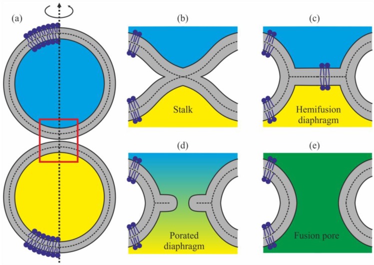
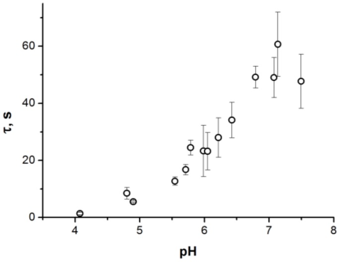
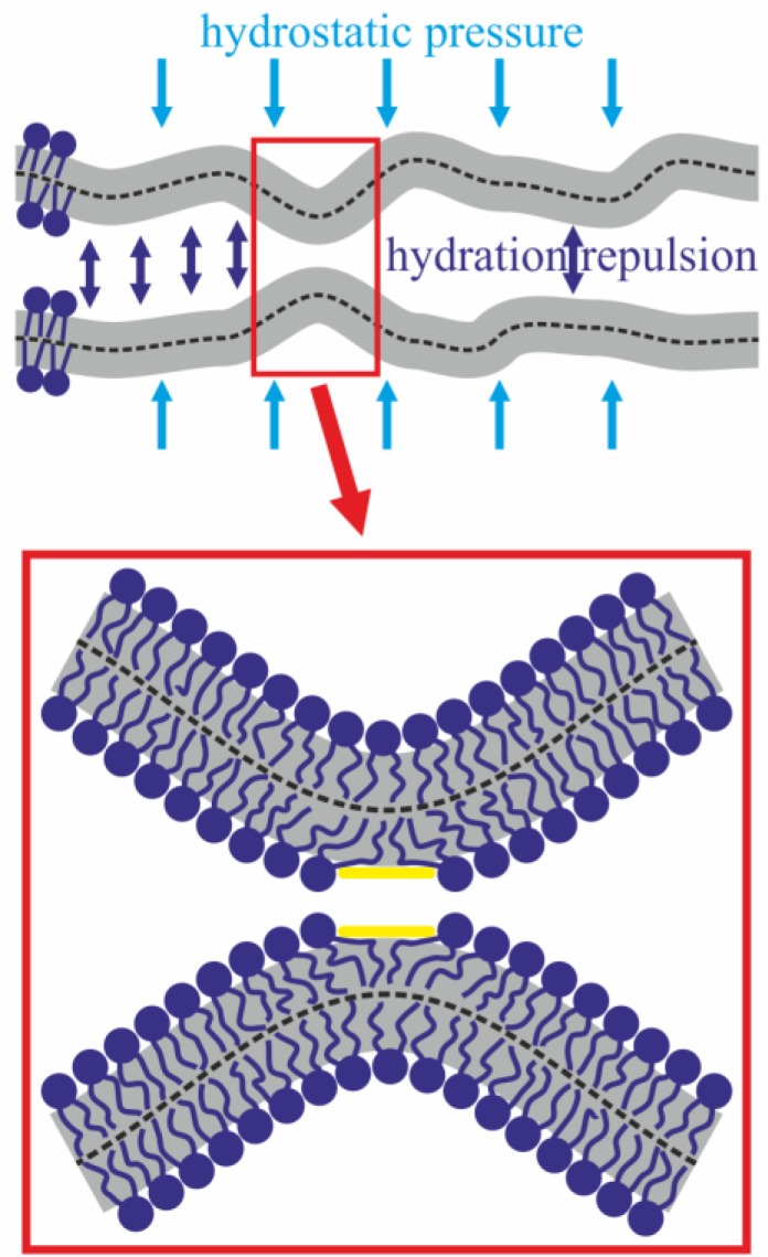
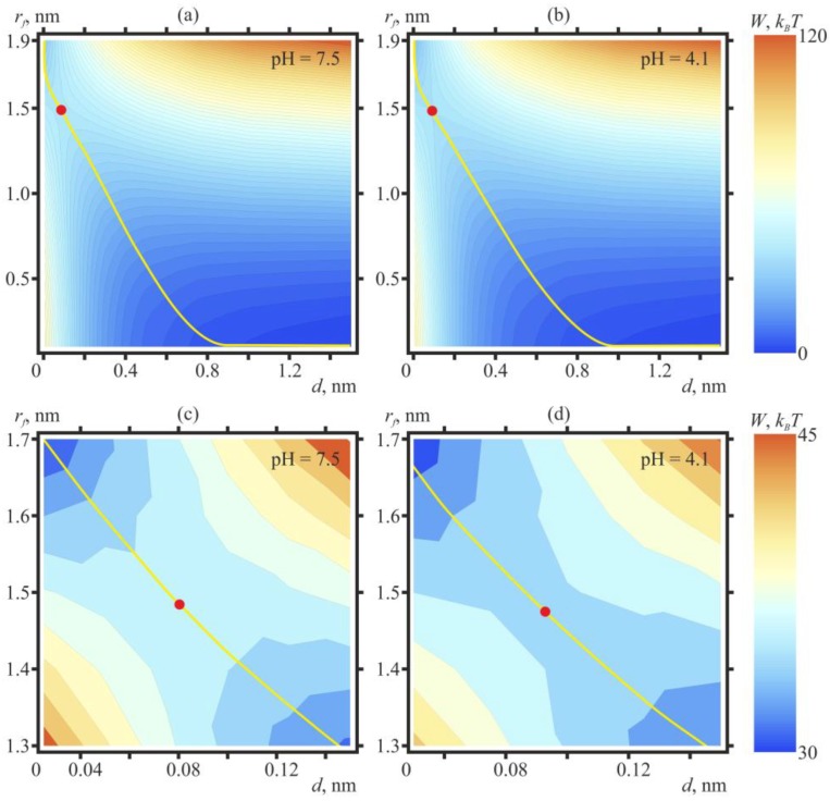
Membrane fusion mediates multiple vital processes in cell life. Specialized proteins mediate the fusion process, and a substantial part of their energy is used for topological rearrangement of the membrane lipid matrix. Therefore, the elastic parameters of lipid bilayers are of crucial importance for fusion processes and for determination of the energy barriers that have to be crossed for the process to take place. In the case of fusion of enveloped viruses (e.g., influenza) with endosomal membrane, the interacting membranes are in an acidic environment, which can affect the membrane's mechanical properties. This factor is often neglected in the analysis of virus-induced membrane fusion. In the present work, we demonstrate that even for membranes composed of zwitterionic lipids, changes of the environmental pH in the physiologically relevant range of 4.0 to 7.5 can affect the rate of the membrane fusion notably. Using a continual model, we demonstrated that the key factor defining the height of the energy barrier is the spontaneous curvature of the lipid monolayer. Changes of this parameter are likely to be caused by rearrangements of the polar part of lipid molecules in response to changes of the pH of the aqueous solution bathing the membrane.

Keywords: enveloped virus; influenza; membrane fusion; pH dependence; stalk; theory of elasticity.


Conflict of interest statement

The authors declare no conflict of interest.

Figures

Figure 1
Figure 1
Schematics of a possible trajectory of the membrane fusion process. The membranes are shown in gray, the water volumes surrounded by them in blue and yellow. (a) Convergence of membranes with formation of tight contact. The contact location is designated by the red rectangle. (b) Stalk, a structure, in which the contact monolayers of membranes have already fused, while the distal ones have not yet done so. (c) Hemifusion diaphragm: during stalk expansion, lipids of the distal monolayers are brought into contact, thus forming a bilayer. (d) During diaphragm poration, the originally isolated aqueous compartments start mixing. (e) Fusion pore.
Figure 2
Figure 2
Dependence of the average waiting time for monolayer fusion of model lipid membranes upon pH of the solution. Each point represents a value averaged over 10 measurements. For points at pH = 4.1 and 4.9 the error bars are directed inside the representing circles, as the statistical error for these points is smaller than the size of the circle; thus, the circles appear as gray (partially filled).
Figure 3
Figure 3
Top—Bilayer lipid membranes thermally fluctuate around the equilibrium distance between the monolayers, determined by the applied hydrostatic pressure and hydration repulsion. On the fluctuation-induced bulges facing each other (surrounded by red rectangle), lipids can displace laterally with the formation of hydrophobic spots (shown in yellow) at the expense of the hydration-induced repulsion.
Figure 4
Figure 4
Contour plot (equal energy lines) of W(rf, d), calculated for dioleoylphosphatidylcholine (DOPC):dioleoylphosphatidylethanolamine (DOPE) = 5:1 membranes at pH = 7.5 (a,c) and at pH = 4.1 (b,d). For plots (a,b) blue color corresponds to 0, red—to 120 kBT; the distance between the isolines is 2 kBT. Red circles outline the saddle points of the energy surface, defining the heights of the energy barriers along the optimal trajectories of the fusion process (shown by yellow lines). The energy in the saddle points amounts to the following values: at pH = 7.5 (a)—39.5 kBT; at pH = 4.1 (b)—36 kBT. The plot (c) presents the zoomed vicinity of the saddle point of the plot (a) (pH = 7.5); the plot (d)—vicinity of the saddle point of the plot (b) (pH = 4.1). For plots (c,d) blue color corresponds to 30 kBT, red—to 45 kBT.
Figure 5
Figure 5
Schematic representation of the experimental cell used for investigation of the model bilayer lipid membranes. G is the generator; A1 and A2 are operational amplifiers. Green arrows represent the directions of the movement of the cell parts.

References

    1. Chernomordik L.V., Kozlov M.M. Mechanics of membrane fusion. Nat. Struct. Mol. Biol. 2008;15:675–683. doi: 10.1038/nsmb.1455. - DOI - PMC - PubMed
    1. McNeil P.L., Steinhardt R.A. Plasma membrane disruption: repair, prevention, adaptation. Annu. Rev. Cell Dev. Biol. 2003;19:697–731. doi: 10.1146/annurev.cellbio.19.111301.140101. - DOI - PubMed
    1. Wassarman P.M. Mammalian fertilization. Cell. 1999;96:175–183. doi: 10.1016/S0092-8674(00)80558-9. - DOI - PubMed
    1. Chen E.H., Grote E., Mohler W., Vignery A. Cell-cell fusion. FEBS Lett. 2007;581:2181–2193. doi: 10.1016/j.febslet.2007.03.033. - DOI - PubMed
    1. Mohler W.A., Shemer G., del Campo J.J., Valansi C., Opoku-Serebuoh E., Scranton V., Assaf N., White J.G., Podbilewicz B. The type I membrane protein EFF-1 is essential for developmental cell fusion. Dev. Cell. 2002;2:355–362. doi: 10.1016/S1534-5807(02)00129-6. - DOI - PubMed

LinkOut - more resources