Review
. 2018 Jul 2;217(7):2235-2246.
doi: 10.1083/jcb.201803152. Epub 2018 May 11.

Fueling thought: Management of glycolysis and oxidative phosphorylation in neuronal metabolism

Affiliations
Review

Fueling thought: Management of glycolysis and oxidative phosphorylation in neuronal metabolism

Gary Yellen. J Cell Biol. .

Abstract

The brain's energy demands are remarkable both in their intensity and in their moment-to-moment dynamic range. This perspective considers the evidence for Warburg-like aerobic glycolysis during the transient metabolic response of the brain to acute activation, and it particularly addresses the cellular mechanisms that underlie this metabolic response. The temporary uncoupling between glycolysis and oxidative phosphorylation led to the proposal of an astrocyte-to-neuron lactate shuttle whereby during stimulation, lactate produced by increased glycolysis in astrocytes is taken up by neurons as their primary energy source. However, direct evidence for this idea is lacking, and evidence rather supports that neurons have the capacity to increase their own glycolysis in response to stimulation; furthermore, neurons may export rather than import lactate in response to stimulation. The possible cellular mechanisms for invoking metabolic resupply of energy in neurons are also discussed, in particular the roles of feedback signaling via adenosine diphosphate and feedforward signaling by calcium ions.


Figures

Figure 1.
Figure 1.
Expression of ample levels of glycolytic (and related) enzymes in both astrocytes and neurons of the central nervous system. Left: Transcriptional expression (from quantitative RNA sequencing) of the glycolytic enzymes and the cytosolic dehydrogenases associated with the NADH shuttles. Data from Zhang et al. (2014) use acutely purified neurons and astrocytes from mouse brain; data from Zeisel et al. (2015) tell a qualitatively similar story. Right: Proteomics data (Sharma et al., 2015) from cultured neurons and astrocytes confirm the general pattern from the transcriptomics data. The data plotted are for cultured astrocytes and for cultured neurons at day-in-vitro 15. Common names are given at the left, and gene names are shown between the two graphs. The largest proportional deficit in glycolysis is a ∼3.6-fold lower total aldolase in neurons versus astrocytes. Note that in neurons, the expression level differences for LDH isoforms and for MCT isoforms are actually quite small using either measure of expression. The expression level for the cytosolic malate dehydrogenase (MDH1) is substantially higher than for cytosolic glycerol-P-dehydrogenase (GPD1), consistent with the dominance of the MAS over the glycerol–phosphate shuttle. FPKM, fragments per kilobase million; IBAQ, integrity-based absolute quantitation.
Figure 2.
Figure 2.
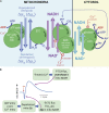
Relationships of some major bioenergetic pools in the neuronal cytosol and mitochondria and their regulation. (A) This schematic shows key bioenergetic processes as waterwheels or gears as well as their interaction with three major bioenergetic reservoirs: the membrane potential across the mitochondrial inner membrane (Δψm) and the related electrochemical gradient for protons across the mitochondrial inner membrane (ΔμH); the mitochondrial NADH/NAD+ redox; and the cytosolic NADH/NAD+ redox. Both in the mitochondrion and cytosol, ADP and Ca2+ are the proximal signals for metabolic demand. As explained in the text, both ADP and Ca2+ entry into mitochondria contribute to mitochondrial depolarization; this allows the ETC to consume mitochondrial NADH and O2 and to restore the mitochondrial ΔμH. (B) The impact of neuronal stimulation on these pathways as measured experimentally by monitoring NAD(P)H autofluorescence is diagrammed. The consumption of mitochondrial NADH leads to the dip in mitochondrial NADH. The increase in mitochondrial [Ca2+] stimulates the TCA cycle and produces an overshoot in NADH production. In the cytosol, NADH/NAD+ redox is controlled by glycolysis (which increases NADH), the MAS, which decreases NADH, and the LDH reaction, which can run in either direction.

References

    1. Ashrafi G., and Ryan T.A.. 2017. Glucose metabolism in nerve terminals. Curr. Opin. Neurobiol. 45:156–161. 10.1016/j.conb.2017.03.007 - DOI - PMC - PubMed
    1. Ashrafi G., Wu Z., Farrell R.J., and Ryan T.A.. 2017. GLUT4 mobilization supports energetic demands of active synapses. Neuron. 93:606–615. 10.1016/j.neuron.2016.12.020 - DOI - PMC - PubMed
    1. Attwell D., and Laughlin S.B.. 2001. An energy budget for signaling in the grey matter of the brain. J. Cereb. Blood Flow Metab. 21:1133–1145. 10.1097/00004647-200110000-00001 - DOI - PubMed
    1. Bak L.K., and Schousboe A.. 2017. Misconceptions regarding basic thermodynamics and enzyme kinetics have led to erroneous conclusions regarding the metabolic importance of lactate dehydrogenase isoenzyme expression. J. Neurosci. Res. 95:2098–2102. 10.1002/jnr.23994 - DOI - PubMed
    1. Bak L.K., and Walls A.B.. 2018. CrossTalk opposing view: lack of evidence supporting an astrocyte-to-neuron lactate shuttle coupling neuronal activity to glucose utilisation in the brain. J. Physiol. 596:351–353. 10.1113/JP274945 - DOI - PMC - PubMed

Publication types