. 2018 May 1;9(5):571.
doi: 10.1038/s41419-018-0632-8.

Caspase-mediated cleavage of the centrosomal proteins during apoptosis

Affiliations

Caspase-mediated cleavage of the centrosomal proteins during apoptosis

Mi Young Seo et al. Cell Death Dis. .

Abstract

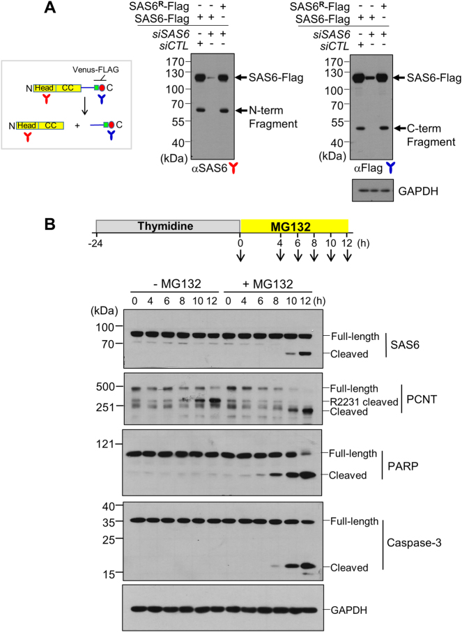
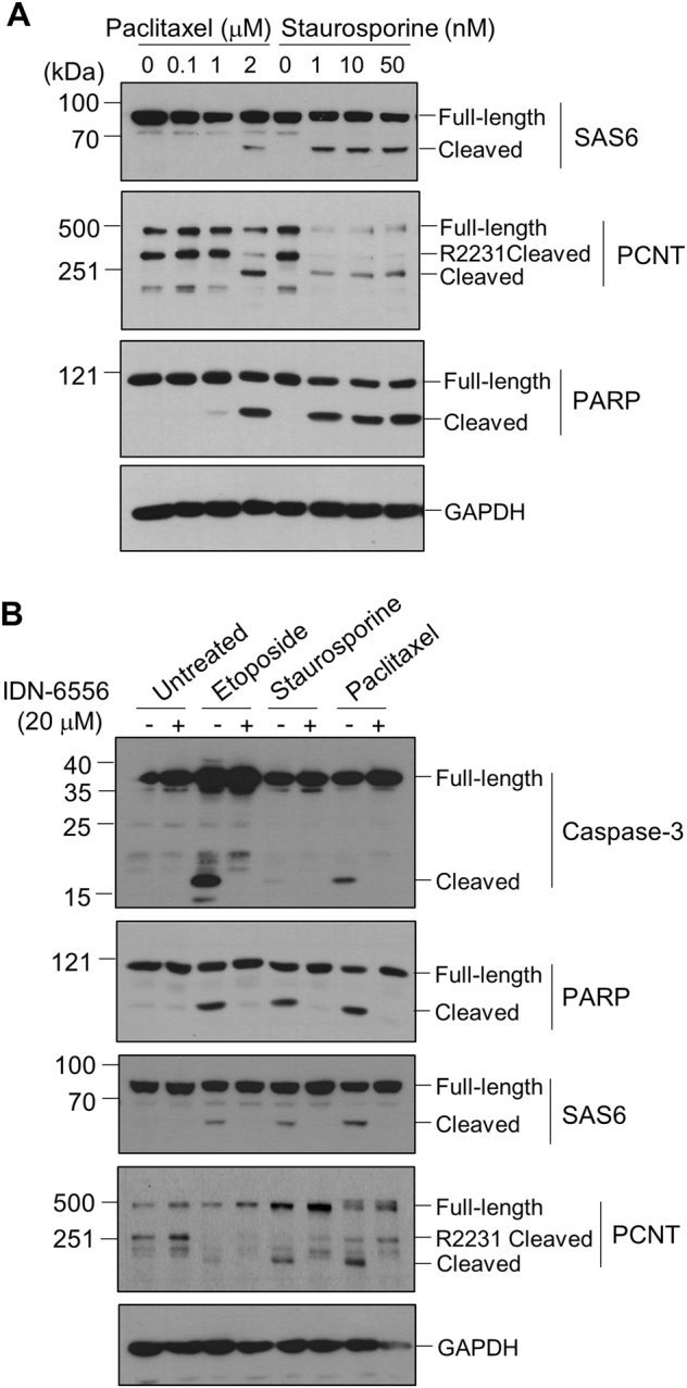
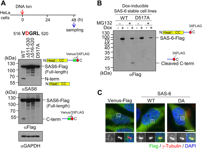
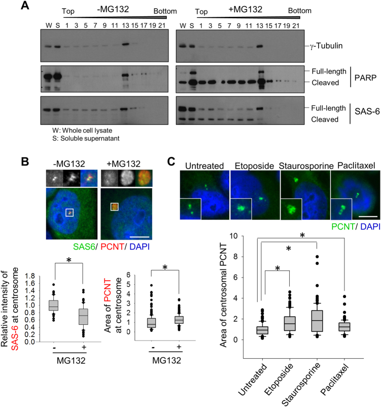
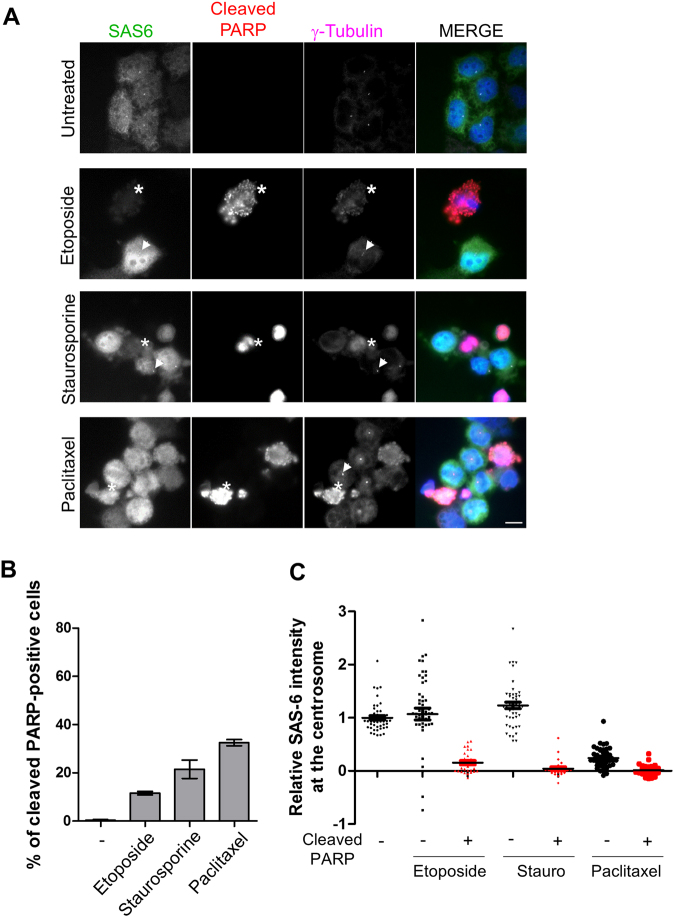
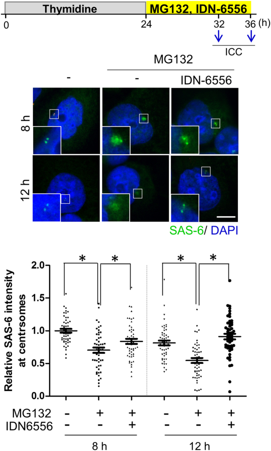
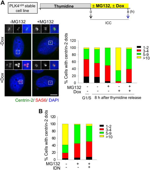
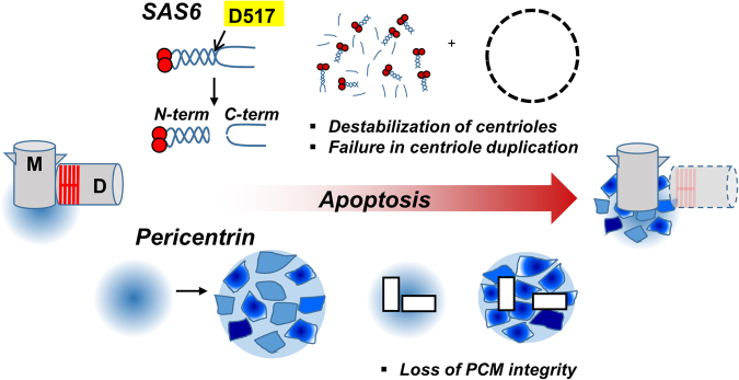
The centrosome is the major microtubule-organizing center and plays important roles in intracellular transport, cellular morphology, and motility. In mitotic cells, centrosomes function as spindle poles to pull a set of chromosomes into daughter cells. In quiescent cells, primary cilia are originated from the centrosomes. Given its involvement in various cellular processes, it is little surprising that the organelle would also participate in apoptotic events. However, it remains elusive how the centrosome changes in structure and organization during apoptosis. Apoptosis, a programmed cell death, is required for homeostatic tissue maintenance, embryonic development, stress responses, etc. Activation of caspases generates a cascade of apoptotic pathways, explaining much of what happens during apoptosis. Here, we report the proteolytic cleavage of selected centrosomal proteins in apoptotic cells. SAS-6, a cartwheel component of centrioles, was specifically cleaved at the border of the coiled-coil domain and the disordered C-terminus. Pericentrin, a scaffold of pericentriolar material, was also cleaved during apoptosis. These cleavages were efficiently blocked by the caspase inhibitors. We propose that the caspase-dependent proteolysis of the centrosomal proteins may destabilize the configuration of a centrosome. Loss of centrosomes may be required for the formation of apoptotic microtubule networks, which are essential for apoptotic fragmentation. This work demonstrates the first centrosomal targets by caspases during apoptosis.


Conflict of interest statement

The authors declare that they have no conflict of interest.

Figures

Fig. 1
Fig. 1. Detection of a specific cleavage of SAS-6.
a HeLa cells were transfected with pSAS-6-Venus-Flag, along with siCTL or siSAS-6. SAS-6R-Flag includes silent mutations resistant to siSAS-6. Forty-eight hours later, the cells were subjected to immunoblot analyses with antibodies specific to SAS-6 and Flag. b HeLa cells were treated with thymidine for 24 h and transferred into a fresh medium with or without MG132. The cells were harvested at the indicated time points and subjected to immunoblot analyses with antibodies specific to SAS-6, pericentrin (PCNT), PARP-1, caspase-3, and GAPDH. Pericentrin cleavage normally occurs during mitotic exit and the cleavage band was labeled as R2231 cleaved
Fig. 2
Fig. 2. Caspase-dependent cleavage of SAS-6 and pericentrin during apoptosis.
a HeLa cells were treated with paclitaxel or staurosporine for 24 h and subjected to immunoblot analyses with antibodies specific to SAS-6, pericentrin (PCNT), PARP-1, and GAPDH. b HeLa cells were treated with etoposide, staurosporine, or paclitaxel in the presence of IDN-6566 for 24 h. The cells were subjected to immunoblot analyses with indicated antibodies
Fig. 3
Fig. 3. Identification of the specific cleavage site of SAS-6.
a Immunoblot analysis was performed with HeLa cells expressing the wild type, the truncated (Δ511–530 and Δ516–520), or the substituted (D517A) mutant of SAS-6-Flag. The ectopic SAS-6 proteins were detected with antibodies specific to SAS-6 and Flag. b SAS-6-stable cell lines of the wild type and the D517A mutant. It is of note that the recombinant SAS-6 expresses a 3xFLAG at its C-terminus with the Venus tag removed. While the wild type generated a cleaved C-terminus fragment, the D517A mutant was resistant to the cleavage in the presence of MG132. c Immunostaining analysis of the wild type and the D517A mutant of ectopic SAS-6 proteins was performed with the Flag antibody. Scale bar, 10 μm
Fig. 4
Fig. 4. Effects of apoptotic agents on the centrosomal SAS-6 and pericentrin.
a HeLa cells were treated with thymidine and transferred into a medium with MG132. Ten hours later, the cell lysates were fractionated with sucrose-gradient ultracentrifugation and subjected to immunoblot analyses with antibodies specific to γ-tubulin, PARP-1, and SAS-6. b The G2 phase HeLa cells were treated with MG132 for 8 h and coimmunostained with antibodies specific to SAS-6 (green) and pericentrin (red). DNA was visualized with DAPI (blue). Scale bar, 10 μm. The centriolar intensity of SAS-6 and centrosomal area of pericentrin were measured and presented with box and whisker plots. c HeLa cells were treated with etoposide, staurosporine, or paclitaxel for 24 h and immunostained with the pericentrin antibody. Greater than 100 centrosomes per experimental group were analyzed in two independent experiments. *P < 0.05
Fig. 5
Fig. 5. Reduction of the centriolar SAS-6 levels in cells undergoing apoptosis.
a HeLa cells were treated with etoposide, staurosporine, or paclitaxel for 24 h and subjected to coimmunostaining with antibodies specific to SAS-6 (green), cleaved PARP-1 (red), and γ-tubulin (magenta). Asterisks indicate the cleaved PARP-positive cells whose SAS-6 intensities were significantly reduced. The arrowheads represent the cleaved PARP-negative cells with the remaining SAS-6 signals at the centrosomes. b Proportions of the cleaved PARP-1-positive cells were determined. c The centriolar intensities of SAS-6 were measured in cleaved PARP-1-negative or PARP-1-positive cells and analyzed with a scatter plot. Greater than 100 centrosomes per experimental group were analyzed in two independent experiments. *P < 0.05
Fig. 6
Fig. 6. Effects of IDN-6556 on the centriolar SAS-6 levels in cells undergoing apoptosis.
HeLa cells were treated with thymidine for 24 h, released into a fresh medium with MG132 and IDN-6566, and cultured for 8 or 12 h. The cells were immunostained with the SAS-6 (green) antibody. DNA was visualized with DAPI (blue). Scale bar, 10 μm. The centriolar intensity of SAS-6 was measured and analyzed with a scatter plot. Greater than 100 centrosomes per experimental group were analyzed in two independent experiments. *P < 0.05
Fig. 7
Fig. 7. Inhibition of the proteasome activity affects the efficiency of centriole duplication in PLK4Δ24 cells.
a HeLa cells were treated with thymidine for 24 h, and transferred into the medium with MG132. Expression of PLK4Δ24 was induced with doxycycline at the same time. At the indicated time points, the cells were coimmunostained with antibodies specific to centrin-2 (green) and SAS-6 (red). DNA was stained with DAPI (blue). Scale bar, 10 μm. The number of centrioles per cell were determined with the centrin-2 signals. b The PLK4Δ24-expressing cells were treated with IDN-6556 and their centriole numbers were counted. Greater than 100 cells per experimental group were analyzed in two independent experiments
Fig. 8
Fig. 8. Model.
During apoptosis, selected centrosomal proteins, such as SAS-6 and pericentrin, are cleaved by caspases. The specific cleavage of SAS-6 might destabilize the procentrioles and prevent a new centriole assembly. Caspase-dependent cleavage of pericentrin might attribute to loss of PCM integrity

References

    1. Dammermann A, et al. Centriole assembly requires both centriolar and pericentriolar material proteins. Dev. Cell. 2004;7:815–829. doi: 10.1016/j.devcel.2004.10.015. - DOI - PubMed
    1. Leidel S, Delattre M, Cerutti L, Baumer K, Gonczy P. SAS-6 defines a protein family required for centrosome duplication in C. elegans and in human cells. Nat. Cell Biol. 2005;7:115–125. doi: 10.1038/ncb1220. - DOI - PubMed
    1. Nakazawa Y, Hiraki M, Kamiya R, Hirono M. SAS-6 is a cartwheel protein that establishes the 9-fold symmetry of the centriole. Curr. Biol. 2007;17:2169–2174. doi: 10.1016/j.cub.2007.11.046. - DOI - PubMed
    1. van Breugel M, et al. Structures of SAS-6 suggest its organization in centrioles. Science. 2011;331:1196–1199. doi: 10.1126/science.1199325. - DOI - PubMed
    1. Kitagawa D, et al. Structural basis of the 9-fold symmetry of centrioles. Cell. 2011;144:364–375. doi: 10.1016/j.cell.2011.01.008. - DOI - PMC - PubMed

Publication types

LinkOut - more resources