. 2019 Jan;76(1):163-172.
doi: 10.1002/cm.21453. Epub 2018 Sep 10.

A requirement for septins and the autophagy receptor p62 in the proliferation of intracellular Shigella

Affiliations

A requirement for septins and the autophagy receptor p62 in the proliferation of intracellular Shigella

Damián Lobato-Márquez et al. Cytoskeleton (Hoboken). 2019 Jan.

Abstract

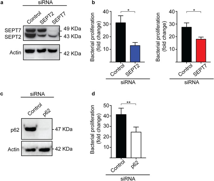
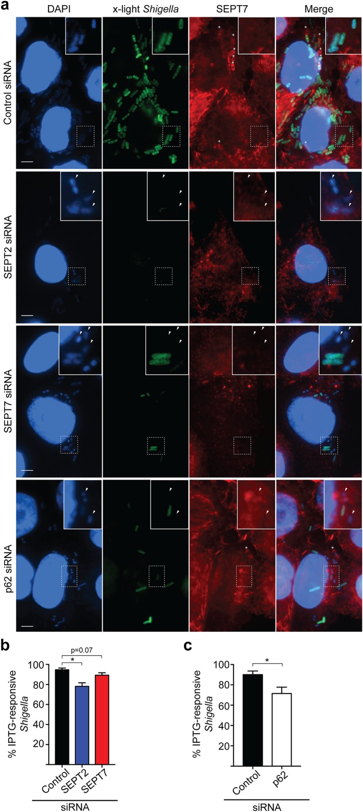
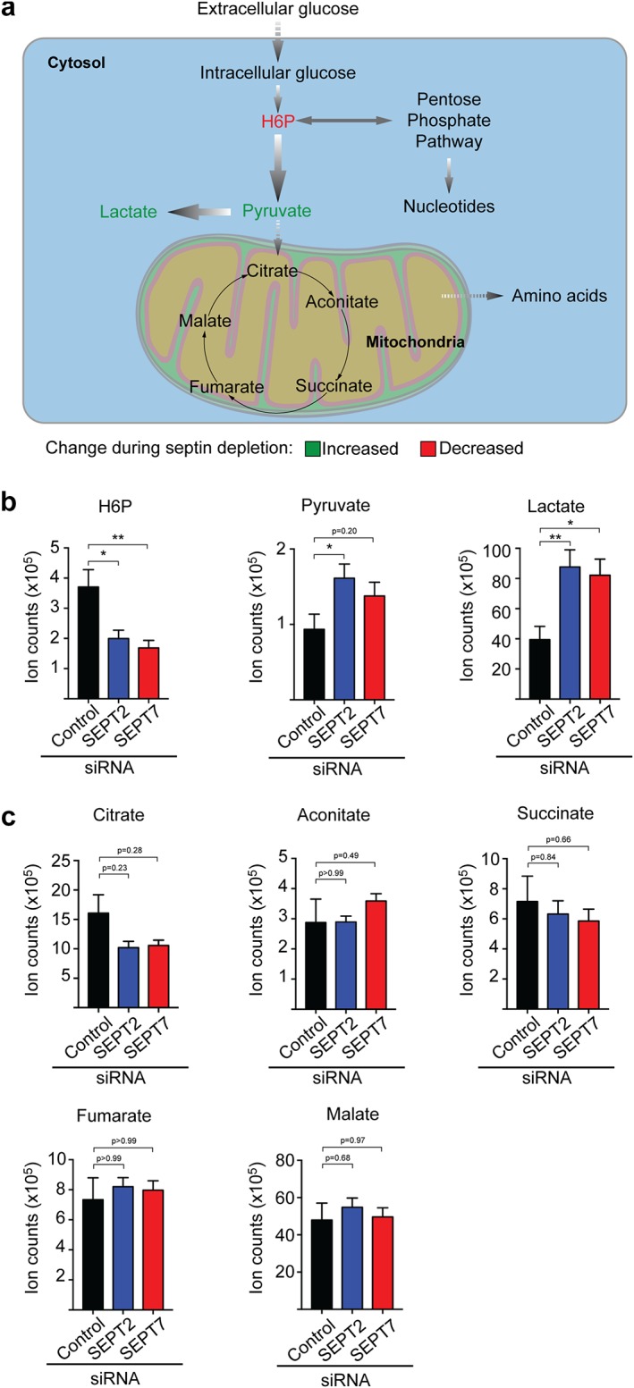
Shigella flexneri, a Gram-negative enteroinvasive pathogen, causes inflammatory destruction of the human intestinal epithelium. During infection of epithelial cells, Shigella escape from the phagosome to the cytosol, where they reroute host cell glycolysis to obtain nutrients for proliferation. Septins, a poorly understood component of the cytoskeleton, can entrap cytosolic Shigella targeted to autophagy in cage-like structures to restrict bacterial proliferation. Although bacterial entrapment by septin caging has been the subject of intense investigation, the role of septins and the autophagy machinery in the proliferation of noncaged Shigella is mostly unknown. Here, we found that intracellular Shigella fail to efficiently proliferate in SEPT2-, SEPT7-, or p62/SQSTM1-depleted cells. Consistent with a failure to proliferate, single cell analysis of bacteria not entrapped in septin cages showed that the number of metabolically active Shigella in septin- or p62-depleted cells is reduced. Targeted metabolomic analysis revealed that host cell glycolysis is dysregulated in septin-depleted cells, suggesting a key role for septins in modulation of glycolysis. Together, these results suggest that septins and the autophagy machinery may regulate metabolic pathways that promote the proliferation of intracellular Shigella not entrapped in septin cages.

Keywords: Shigella; autophagy; cytoskeleton; metabolism; septin.


Figures

Figure 1
Figure 1
SEPT2, SEPT7 and p62 promote the proliferation of intracellular Shigella. (a) HeLa cells were treated with control, SEPT2, or SEPT7 siRNA sequences for 72 h. Whole‐cell lysates were immunoblotted for SEPT2 and SEPT7 to show the efficiency of depletion. Actin was used as a loading control. (b) siRNA‐treated cells were infected with S. flexneri str. M90T for 1 h 40 min or 4 h 40 min, then lysed and plated on LB‐agar plates. Graphs represent mean fold replication ratio 4 h 40 min/1 h 40 min ± SEM from four independent experiments. (c) HeLa cells were treated with control or p62 siRNA sequences for 72 h. Whole‐cell lysates were immunoblotted for p62 to show the efficiency of depletion. Actin was used as a loading control. (d) siRNA‐treated cells were infected with S. flexneri str. M90T as in (b) [Color figure can be viewed at wileyonlinelibrary.com]
Figure 2
Figure 2
SEPT2, SEPT7, and p62 promote the metabolic activity of intracellular Shigella not entrapped in septin cages. (a) siRNA‐treated HeLa cells were infected with x‐light S. flexneri for 4 h 10 min, before being exposed to IPTG during 30 min prior to fixation. Samples were labeled for SEPT7 antibody to exclude septin caged bacteria from analysis. Arrowheads indicate metabolically inactive bacteria. *, septin cages. Scale bars = 5 μm. (b,c) HeLa cells were infected with x‐light S. flexneri as described in (a) and then the number of IPTG‐responsive intracellular bacteria was quantified in SEPT2‐, SEPT7‐ (b), or p62‐depleted cells (c). Graph represents mean % IPTG‐responsive bacteria ± SEM from four independent experiments. *p < .05 as analyzed by one‐way ANOVA (b) or Student's t test (c) [Color figure can be viewed at wileyonlinelibrary.com]
Figure 3
Figure 3
SEPT2 and SEPT7 regulate glycolysis in human epithelial cells. (a) Scheme of the primary metabolic routes in human epithelial cells. Arrows indicate the flux of each metabolic pathway. H6P, hexose‐6‐phosphate. (b,c) HeLa cells were treated with control, SEPT2, or SEPT7 siRNA sequences for 72 h. Cell lysates were prepared for LC–MS AMRT analysis. (b) Individual quantifications for indicated glycolytic metabolites, (c) individual quantifications for indicated TCA cycle metabolites. Graphs represent mean ± SEM of normalized ion counts from three independent experiments done in duplicate. *p < .05, **p < .001 as analyzed by one‐way ANOVA [Color figure can be viewed at wileyonlinelibrary.com]

References

    1. Abu Kwaik, Y. , & Bumann, D. (2015). Host delivery of favorite meals for intracellular pathogens. PLoS Pathogens, 11(6), e1004866. - PMC - PubMed
    1. Araiza‐Olivera, D. , Chiquete‐Felix, N. , Rosas‐Lemus, M. , Sampedro, J. G. , Peña, A. , Mujica, A. , & Uribe‐Carvajal, S. (2013). A glycolytic metabolon in Saccharomyces cerevisiae is stabilized by F‐actin. FEBS Journal, 280(16), 3887–3905. - PubMed
    1. Barve, G. , Sridhar, S. , Aher, A. , Sahani, M. H. , Chinchwadkar, S. , Singh, S. , … Manjithaya, R. (2018). Septins are involved at the early stages of macroautophagy in S. cerevisiae . Journal of Cell Science, 131(4). pii: jcs209098. 10.1242/jcs.209098. - DOI - PMC - PubMed
    1. Choy, A. , Dancourt, J. , Mugo, B. , O'Connor, T. J. , Isberg, R. R. , Melia, T. J. , & Roy, C. R. (2012). The Legionella effector RavZ inhibits host autophagy through irreversible Atg8 deconjugation. Science, 338(6110), 1072–1076. - PMC - PubMed
    1. Chung, H. , & Baker, S. (2018). Out of Asia: The independent rise and global spread of fluoroquinolone‐resistant Shigella . Microbial Genomics, 4(4), - PMC - PubMed

Publication types