. 2018 Aug:173:109-112.
doi: 10.1016/j.exer.2018.05.002. Epub 2018 May 9.

Epitope mapping of commercial antibodies that detect myocilin

Affiliations

Epitope mapping of commercial antibodies that detect myocilin

Athéna C Patterson-Orazem et al. Exp Eye Res. 2018 Aug.

Abstract

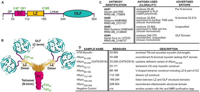
The presence of myocilin is often used in the process of validating trabecular meshwork (TM) cells and eye tissues, but the antibody reagents used for detection are poorly characterized. Indeed, for over a century, researchers have been using antibodies to track proteins of interest in a variety of biological contexts, but many antibodies remain ill-defined at the molecular level and in their target epitope. Such issues have prompted efforts from major funding agencies to validate reagents and combat reproducibility issues across biomedical sciences. Here we characterize the epitopes recognized by four commercial myocilin antibodies, aided by structurally and biochemically characterized myocilin fragments. All four antibodies recognize enriched myocilin secreted from human TM cell media. The detection of myocilin fragments by ELISA and Western blot reveal a variety of epitopes across the myocilin polypeptide chain. A more precise understanding of myocilin antibody targets, including conformational specificity, should aid the community in standardizing protocols across laboratories and in turn, lead to a better understanding of eye physiology and disease.

Keywords: Antibodies; Antigen; ELISA; Epitopes; Glaucoma; Myocilin; Trabecular meshwork; Western blot.


Figures

Fig. 1.
Fig. 1.
Introduction to myocilin structure, commercial antibodies, and protein constructs tested. (A) Gene structure depicting the domains of myocilin, including signal peptide (SP), location of key cysteine residues (C47, C61 and C185) and its coiled-coil (CC), leucine zipper (LZ) and olfactomedin (OLF) domains. (B) Myocilin quaternary structure based on solution X-ray scattering, X-ray crystallography and chemical cross-linking experiments. (C) Identifying information for commercial myocilin antibodies selected for this study, including antigen used for antibody development, clonality, and currently advertised epitope. (D) Myocilin constructs used in this study represent a combination of shorter and longer myocilin fragments. Each potential epitope for antibodies is represented at least twice.
Fig. 2.
Fig. 2.
Epitope mapping of commercial antibodies reveals four distinct epitopes. (A,D,G,J) Western blots demonstrate binding of antibodies against cell lysates containing recombinant myocilin fragments under denaturing conditions. Manufacturers’ recommended primary antibody concentrations are indicated with an asterisk, and standards (PageRuler™ Plus, Thermo Scientific) are indicated in kilodaltons (kDa). Note that the size of recombinant constructs is affected by expression tags, including maltose binding protein (~45 kDa) for rMyoc33–226 and rMyoc228–504 and hexahistidine/S-tags for the remaining constructs (~6 kDa). (B,E,H,K) All antibodies tested recognize myocilin enriched from hTM cell-conditioned medium in Western blots. ELISA conducted against clarified cell lysates (C,I) and purified protein (F,L) demonstrate antibody binding to recombinant myocilin constructs in native conditions. (M) Schematic representation of target regions of the four commercial antibodies on myocilin.

References

    1. Anderssohn AM, Cox K, O’Malley K, Dees S, Hosseini M, Boren L, Wagner A, Bradley JM, Kelley MJ, Acott TS, 2011. Molecular chaperone function for myocilin. Invest Ophth Vis Sci 52, 7548–7555. - PMC - PubMed
    1. Begley CG, Ellis LM, 2012. Drug development: Raise standards for preclinical cancer research. Nature 483, 531–533. - PubMed
    1. Bordeaux J, Welsh A, Agarwal S, Killiam E, Baquero M, Hanna J, Anagnostou V, Rimm D, 2010. Antibody validation. Biotechniques 48, 197–209. - PMC - PubMed
    1. Bradbury A, Pluckthun A, 2015a. Reproducibility: Standardize antibodies used in research. Nature 518, 27–29. - PubMed
    1. Bradbury AR, Pluckthun A, 2015b. Getting to reproducible antibodies: the rationale for sequenced recombinant characterized reagents. Protein Eng Des Sel 28, 303–305. - PubMed

Publication types