. 2018 Aug 15;440(2):99-112.
doi: 10.1016/j.ydbio.2018.05.006. Epub 2018 May 9.

The Misshapen kinase regulates the size and stability of the germline ring canals in the Drosophila egg chamber

Affiliations

The Misshapen kinase regulates the size and stability of the germline ring canals in the Drosophila egg chamber

Ashley Kline et al. Dev Biol. .

Abstract

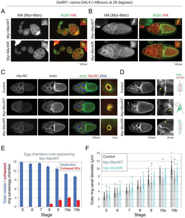
Intercellular bridges are conserved structures that allow neighboring cells to exchange cytoplasmic material; defects in intercellular bridges can lead to infertility in many organisms. Here, we use the Drosophila egg chamber to study the mechanisms that regulate intercellular bridges. Within the developing egg chamber, the germ cells (15 nurse cells and 1 oocyte) are connected to each other through intercellular bridges called ring canals, which expand over the course of oogenesis to support the transfer of materials from the nurse cells to the oocyte. The ring canals are enriched in actin and actin binding proteins, and many proteins have been identified that localize to the germline ring canals and control their expansion and stability. Here, we demonstrate a novel role for the Ste20 family kinase, Misshapen (Msn), in regulation of the size of the germline ring canals. Msn localizes to ring canals throughout most of oogenesis, and depletion of Msn led to the formation of larger ring canals. Over-expression of Msn decreased ring canal diameter, and expression of a membrane tethered form of Msn caused ring canal detachment and nurse cell fusion. Altering the levels or localization of Msn also led to changes in the actin cytoskeleton and altered the localization of E-cadherin, which suggests that Msn could be indirectly limiting ring canal size by altering the structure or dynamics of the actin cytoskeleton and/or adherens junctions.

Keywords: Drosophila; Egg chamber; Misshapen (Msn); Oogenesis; Ring canal.


Figures

Figure 1
Figure 1. Misshapen localizes to the germline ring canals and nurse cell membranes throughout oogenesis
(A) Schematic of the germarium and inner and outer rim structure of the ring canals. (B–D) Single plane confocal images of egg chambers expressing a MsnYFP protein trap. Samples were stained with an anti-GFP antibody, phalloidin, and DAPI. Arrowheads point to MsnYFP on nurse cell membranes. (E) Maximum intensity projection of confocal images of egg chambers stained with an anti-GFP antibody, phalloidin, and DAPI. (F) Single plane confocal images of MsnYFP-expressing egg chambers stained with an Hts-RC antibody and DAPI. Boxes indicate magnified regions.
Figure 2
Figure 2. Depletion of Misshapen increases ring canal diameter and causes ring canal collapse
Depletion of Misshapen beginning at stage 2 of oogenesis was performed using matαTub-GAL4. (A) Images of stage 10B control and msn-RNAi egg chambers. (B) Scatter plot showing the outer diameter of individual ring canals connecting nurse cells in control and msn-RNAi egg chambers at each stage. Bars indicate the average outer diameter at each stage. n = 33–101 ring canals/stage for each condition. Asterisks indicate significant difference compared to control (p<0.05, 2-tailed t-test). (C) Average number of ring canals per egg chamber that have a clear lumen or were collapsed. n=6–12 egg chambers/stage. Error bars are SEM. Images of stage 10B control and msn-RNAi egg chambers stained with antibodies against (D) Kelch or (E) phospho-tyrosine. Arrows in (D,E) point to collapsed ring canals. Images in E are maximum intensity projections. Boxes in A, D, and E indicate magnified regions shown in panels to the right. Graphs in A,D,E show the average fluorescence intensity of Hts-RC (n=25 for control, n=19 for msn-RNAi), Kelch (n=20 for each condition), or pTyr (n=12 for control, n=18 for msn-RNAi) staining in ring canals of stage 10 control and msn-RNAi egg chambers. Error bars are SEM. Average full width at half maximum +/− SEM is shown for each stain in each condition. Asterisks indicate significant difference compared to control (p<0.05, 2-tailed t-test).
Figure 3
Figure 3. Over-expression of Misshapen decreases ring canal diameter
(A,B) Fluorescence images of stage 6 and stage 10 HA-MsnWT or HA-MsnKR expressing egg chambers stained with an HA antibody. Boxes indicate regions of magnification shown to the right. The brightness/contrast settings were altered from the larger panels to better visualize ring canal localization. (C) Western blot of whole ovary lysate from control, HA-MsnWT, or HA-MsnKR expressing flies. (D) Fluorescence images of stage 10 control, HA-MsnWT, or HA-MsnKR expressing egg chambers. Boxes indicate regions of magnification shown to the right. (E) Scatter plot showing the outer diameter of individual ring canals connecting nurse cells in control, HA-MsnWT, or HA-MsnKR expressing egg chambers. Bars indicate the average outer diameter at each stage. n = 104–165 ring canals/stage for each condition. Asterisks indicate significant difference compared to control (p<0.05, 2-tailed t-test). All crosses were set up with the maternal triple driver, MTD-GAL4.
Figure 4
Figure 4. Expression of a membrane-tethered form of Misshapen leads to defects in ring canal stability and nurse cell fusion
(A,B) Fluorescence images of egg chambers expressing Myr-HA-MsnWT or Myr-HA-MsnKR stained with an HA antibody. In addition to the ring canals and nurse cell membranes, Myr-HA-MsnWT and Myr-HA-MsnKR localize to punctae in the nurse cells and oocyte (arrowhead). Arrow in (B) points to a collapsed ring canal in a stage 10 egg chamber expressing Myr-HA-MsnWT. Boxes indicate regions of magnification. For magnified regions, scale bars are 2μm. (C,D) Fluorescence images of stage 10 control, Myr-HA-MsnWT, or Myr-HA-MsnKR expressing egg chambers. Boxes indicate regions of magnification in panels on the right. White arrowhead in (D) points to large actin bundle. Yellow arrowheads point to actin structures shown in the cartoons to the right. (E) Average number of visible and collapsed ring canals in egg chambers expressing Myr-HA-MsnWT. n=8–17 egg chambers/stage. Error bars are SEM. (F) Scatter plot showing the outer diameter of individual ring canals connecting nurse cells in control, Myr-HA-MsnWT, and Myr-HA-MsnKR expressing egg chambers. Bars indicate the average outer diameter at each stage. n = 44–159 ring canals/stage for each condition. Crosses were set up with the Gal80ts;nanos-GAL4 driver. Asterisks indicate significant difference compared to control (p<0.05, 2-tailed t-test).
Figure 5
Figure 5. Altering Msn levels leads to changes in E-cadherin localization and defects in border cell migration
Images of stage 9 control and msn-RNAi egg chambers stained with antibodies against (A) E-cadherin or (B) phosphorylated β-catenin (pY142). Crosses were performed with either (A) MTD-GAL4 or (B) matαTub-GAL4. (C) Fluorescence images of stage 9 egg chambers expressing Myr-HA-MsnWT (Gal80ts;nanos-GAL4) and stained with the E-cadherin antibody. Asterisk marks the border cell cluster in (A) and (C), and boxes indicate regions of magnification. (D) Images of stage 10 control and Myr-HA-MsnWT expressing egg chambers (matαTub-GAL4) stained with a FasIII antibody to label the polar cells, which are found in the center of the border cell cluster, and an Hts-RC antibody to label the ring canals. Arrows indicate the position of the border cell cluster. The number of ring canals and degree of border cell migration is indicated for the examples shown. (E) Scatter plot showing the number of ring canals and the percent border cell migration in individual Myr-HA-MsnWT expressing stage 10 egg chambers (n=24). All control egg chambers contained 15 ring canals, and the border cells had migrated 100% (n=30).

References

    1. Bogard N, Lan L, Xu J, Cohen RS. Rab11 maintains connections between germline stem cells and niche cells in the Drosophila ovary. Development. 2007;134:3413–3418. - PubMed
    1. Cai D, Chen SC, Prasad M, He L, Wang X, Choesmel-Cadamuro V, Sawyer JK, Danuser G, Montell DJ. Mechanical feedback through E-cadherin promotes direction sensing during collective cell migration. Cell. 2014;157:1146–1159. - PMC - PubMed
    1. Cobreros-Reguera L, Fernandez-Minan A, Fernandez-Espartero CH, Lopez-Schier H, Gonzalez-Reyes A, Martin-Bermudo MD. The Ste20 kinase misshapen is essential for the invasive behaviour of ovarian epithelial cells in Drosophila. EMBO Rep. 2010;11:943–949. - PMC - PubMed
    1. Cooley L, Verheyen E, Ayers K. chickadee encodes a profilin required for intercellular cytoplasm transport during Drosophila oogenesis. Cell. 1992;69:173–184. - PubMed
    1. Coutelis JB, Ephrussi A. Rab6 mediates membrane organization and determinant localization during Drosophila oogenesis. Development. 2007;134:1419–1430. - PubMed

Publication types

MeSH terms

LinkOut - more resources