. 2019 May:78-79:165-179.
doi: 10.1016/j.matbio.2018.05.002. Epub 2018 May 10.

Prostate tumor cell exosomes containing hyaluronidase Hyal1 stimulate prostate stromal cell motility by engagement of FAK-mediated integrin signaling

Affiliations

Prostate tumor cell exosomes containing hyaluronidase Hyal1 stimulate prostate stromal cell motility by engagement of FAK-mediated integrin signaling

Caitlin O McAtee et al. Matrix Biol. 2019 May.

Abstract

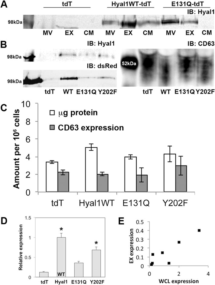
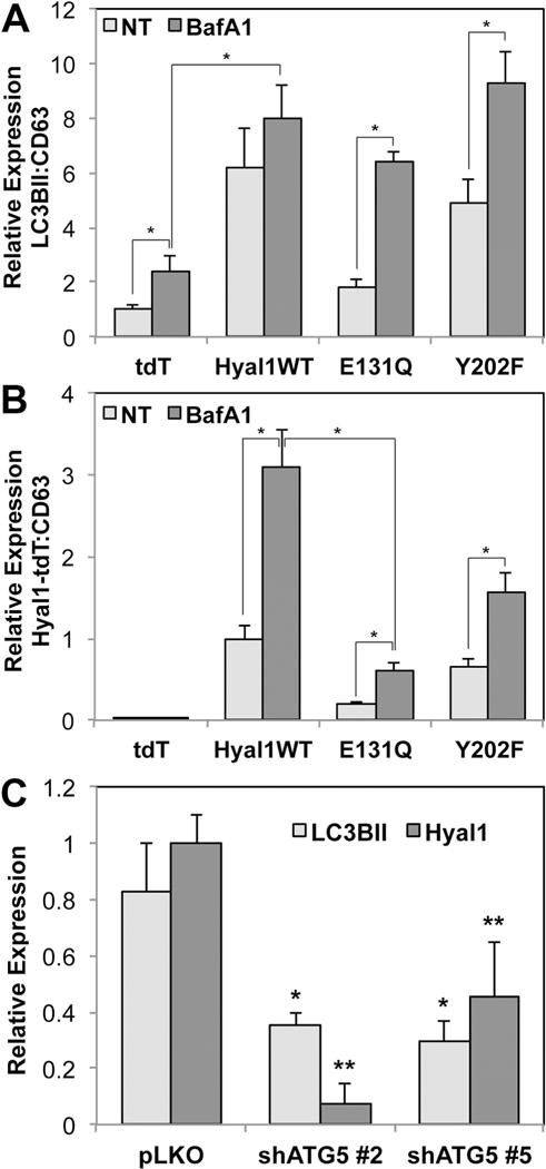
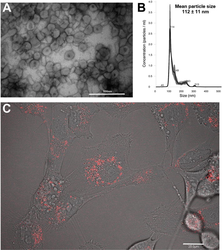
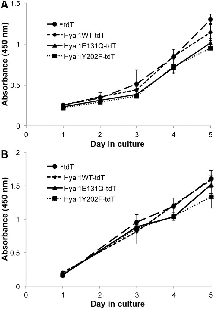
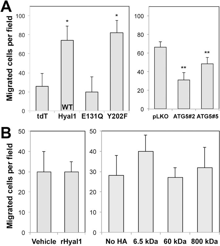
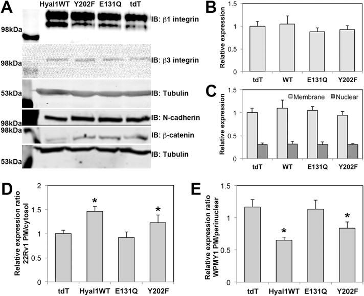
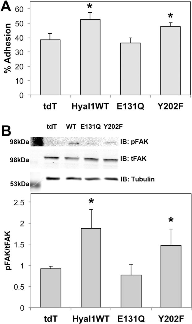
The hyaluronidase Hyal1 is clinically and functionally implicated in prostate cancer progression and metastasis. Elevated Hyal1 accelerates vesicular trafficking in prostate tumor cells, thereby enhancing their metastatic potential in an autocrine manner through increased motility and proliferation. In this report, we found Hyal1 protein is a component of exosomes produced by prostate tumor cell lines overexpressing Hyal1. We investigated the role of exosomally shed Hyal1 in modulating tumor cell autonomous functions and in modifying the behavior of prostate stromal cells. Catalytic activity of Hyal1 was necessary for enrichment of Hyal1 in the exosome fraction, which was associated with increased presence of LC3BII, an autophagic marker, in the exosomes. Hyal1-positive exosome contents were internalized from the culture medium by WPMY-1 prostate stromal fibroblasts. Treatment of prostate stromal cells with tumor exosomes did not affect proliferation, but robustly stimulated their migration in a manner dependent on Hyal1 catalytic activity. Increased motility of exosome-treated stromal cells was accompanied by enhanced adhesion to a type IV collagen matrix, as well as increased FAK phosphorylation and integrin engagement through dynamic membrane residence of β1 integrins. The presence of Hyal1 in tumor-derived exosomes and its ability to impact the behavior of stromal cells suggests cell-cell communication via exosomes is a novel mechanism by which elevated Hyal1 promotes prostate cancer progression.

Keywords: Cell motility; Exosomes; Hyaluronan; Hyaluronidase; Prostate cancer; Stromal-epithelial crosstalk.


Figures

Figure 1
Figure 1. Exosomes secreted by prostate tumor cells contain catalytically active Hyal1
Conditioned media were collected from stable 22Rv1 transfectants expressing tdTomato (tdT, vector control), or the indicated Hyal1 construct, after culturing in exosome-depleted media for 48 hours. Large microvesicle (MV) and exosome (EX) fractions were prepared from conditioned media by differential centrifugation. (A) Equal amounts of protein from each fraction were immunoblotted for Hyal1. CM indicates concentrated media fractions following exosome centrifugation. (B) Exosomal fractions were immunoblotted for Hyal1 (upper left), dsRed (lower left), and CD63 (right). Expected band size is ≈100 kDa for Hyal1-tdT fusion proteins. Expected band ranges from 42–53 kDa for CD63. One representative analysis series is shown from a total of >4 separate exosome preparations. (C) Comparison of exosomal yield per million cells. Protein concentration was determined by BCA assay, CD63 expression was estimated by western blot. Mean ± SEM of three exosomal preparations for each transfectant are presented. No significant differences were observed between these samples. (D) Relative amount of Hyal1 in the exosomal fraction of each cell line was compared by plotting the ratio of dsRed in a region of interest at 100kDa to CD63 in the western analysis. At least four separate preparations of exosomes from each line were analyzed. Mean ± SEM is plotted; *p<0.05 relative to the E131Q-tdT mutant. (E) Pearson’s correlation coefficient was calculated for Hyal1WT-tdT expression in exosomes versus whole cell lysates. ρ=0.88.
Figure 2
Figure 2. Hyal1-containing exosomes are shed partially by an autophagosomal route
(A, B) Hyal1 expression increases exosomal content of LC3BII and Hyal1-containing exosome secretion is stimulated by Bafilomycin A1. Exosome fractions from 22Rv1 cells stably expressing tdT, Hyal1WT-tdT, Hyal1E131Q-tdT, or Hyal1Y202F-tdT were analyzed by western blot for CD63, Hyal1-tdT, and the autophagic marker LC3BII. Triplicates of each blot were quantified to compare relative amounts of LC3BII, normalized to CD63 (A), or Hyal1:CD63 (B) for each of the transfectant lines. Mean ± SEM is plotted; * p<0.05. (C) ATG5 knockdown diminishes exosome shedding, extracellular Hyal1, and Hyal1-positive exosomes, and significantly reduces exosomal Hyal1 content (normalized to CD63). Using PC3 cells selected for vector (pLKO) or ATG5 shRNA (two constructs, #2 and #5), exosomes were analyzed by western blot for CD63, LC3BII, and endogenous Hyal1. Levels of LC3BII and Hyal1 were normalized to CD63 in each preparation. Mean ± SEM is plotted; * p<0.05 for LC3BII expression relative to pLKO; ** p<0.05 for Hyal1 expression relative to pLKO.
Figure 3
Figure 3. Tumor exosome contents are internalized by WPMY-1 prostate stromal fibroblasts
(A) Exosomes were isolated from 22Rv1 Hyal1WT-tdT transfected cell conditioned medium and imaged by TEM to confirm their correct size and morphology (scale bar is 500 nm). (B) Exosomes were characterized by nanoparticle tracking analysis to obtain numbers, average size, and size homogeneity. (C) WPMY-1 cells were seeded overnight on glass bottom tissue culture dishes, exposed to exosomes for 15 minutes, and imaged by fluorescence confocal microscopy to visualize and confirm tdT reporter internalization. Red fluorescent signal was visible in intracellular punctate structures consistent with endosomally internalized vesicles (scale bar is 20 μm, a single subsurface z-section is shown).
Figure 4
Figure 4. Proliferation of tumor and stromal cells is not affected by treatment with exosomes containing Hyal1
Tumor cells (22Rv1, A) or stromal cells (WPMY-1, B) were seeded at equal density in 96 well plates (day “0”). Proliferation was measured daily, or as indicated, by absorbance upon treatment with WST-1. Media were replaced on day 1 and every other day thereafter with RPMI + 5% FBS that contained isolated exosomes from the indicated tumor transfectant lines. Each condition was monitored in quadruplicate wells. Mean ± SEM is plotted for each day. No significant differences were observed among treatments.
Figure 5
Figure 5. Tumor-derived exosomes containing catalytically active Hyal1WT stimulate prostate stromal cell motility
(A) WPMY-1 prostate stromal fibroblasts were treated for 24 h with exosome-depleted media to which aliquots of isolated exosomes from the indicated tumor cell lines were added (22Rv1 exosomal effects shown at left, PC3 effects at right). Migration of exosome-treated or untreated WPMY-1 cells to type IV collagen was compared using a modified Boyden chamber assay. Mean number of cells migrated per field is plotted ± SEM for triplicate wells; * p<0.05 relative to the tdT exosome control treatment; ** p<0.05 relative to the pLKO exosome control treatment. (B) Purified recombinant Hyal1 protein (left) or exogenous HA of different sizes (right) does not affect WPMY-1 motility. Stromal cells were treated with 10 ng/ml rhHyal1 or 10 μg/mL of the indicated size of HA in SFM for 24 h before chemotaxis assay. No significant differences were observed among these treatments.
Figure 6
Figure 6
Exosomes containing active Hyal1 do not affect stromal cell expression of specific motility receptors, but membrane residence of β1 integrin is altered. Stromal cells were treated with exosomes normalized to CD63 expression for 24 hours in SFM. (A) Whole cell lysates of treated cells were analyzed for β1 integrin, β3 integrin, N-cadherin, or β-catenin expression. (B) Plasma membrane enriched fractions were analyzed for N-cadherin expression. (C) Exosome-treated cells were fractionated into membrane and nuclear compartments and analyzed by western blot for β-catenin. (D) Cell surface and cytosolic β1 integrin was imaged by immunofluorescence confocal microscopy of intact and permeabilized 22Rv1 tumor cell transfectant lines cells, respectively (n=10 per condition), quantified in Image J, and plotted as a ratio for surface to cytosol. (E) Cell surface versus cytosolic β1 integrin was imaged by immunofluorescence confocal microscopy of intact and permeabilized exosome-treated stromal cells, respectively (n=20 per condition), quantified in Image J, and plotted as a ratio for surface to perinuclear staining intensity. In B–E, mean ± SEM is plotted; * p<0.05 relative to tdT control transfectants (D) or to stromal cells treated with tdT control exosomes (E).
Figure 7
Figure 7
Active Hyal1-containing exosomes accelerate stromal chemotaxis via increased ECM adhesion and activation of FAK phosphorylation. (A) Exosome-treated or untreated WPMY-1 cells were labeled with calcein-AM and seeded on type IV collagen coated microwell plates for 30 min at 37°C. Non-adherent cells were removed by washing and remaining adherent cells were quantified by fluorescence. (B) Exosome-treated or untreated WPMY-1 cells were seeded on type IV collagen coated plates. After two hours, adherent cells were lysed and equal amounts of protein were analyzed by western blot for phosphorylated and total FAK, which was plotted as a ratio. In both panels, the mean ± SEM for triplicate wells is plotted; *p<0.05 relative to cells treated with tdT control exosomes.

References

    1. Aaltomaa S, Lipponen P, Tammi R, Tammi M, Viitanen J, Kankkunen JP, Kosma VM. Strong Stromal Hyaluronan Expression Is Associated with PSA Recurrence in Local Prostate Cancer. Urol Int. 2002;69(4):266–72. - PubMed
    1. Ekici S, Ayhan A, Kendi S, Ozen H. Determination of prognosis in patients with prostate cancer treated with radical prostatectomy: prognostic value of CD44v6 score. The Journal of urology. 2002;167(5):2037–41. - PubMed
    1. McAtee CO, Barycki JJ, Simpson MA. Emerging roles for hyaluronidase in cancer metastasis and therapy. Advances in cancer research. 2014;123:1–34. - PMC - PubMed
    1. Posey JT, Soloway MS, Ekici S, Sofer M, Civantos F, Duncan RC, Lokeshwar VB. Evaluation of the prognostic potential of hyaluronic acid and hyaluronidase (HYAL1) for prostate cancer. Cancer research. 2003;63(10):2638–44. - PubMed
    1. Simpson MA, Lokeshwar VB. Hyaluronan and hyaluronidase in genitourinary tumors. Front Biosci. 2008;13:5664–80. - PMC - PubMed

Publication types

MeSH terms