. 2018 Jul;136(1):139-152.
doi: 10.1007/s00401-018-1862-7. Epub 2018 May 12.

Convective influx/glymphatic system: tracers injected into the CSF enter and leave the brain along separate periarterial basement membrane pathways

Affiliations

Convective influx/glymphatic system: tracers injected into the CSF enter and leave the brain along separate periarterial basement membrane pathways

Nazira J Albargothy et al. Acta Neuropathol. 2018 Jul.

Abstract

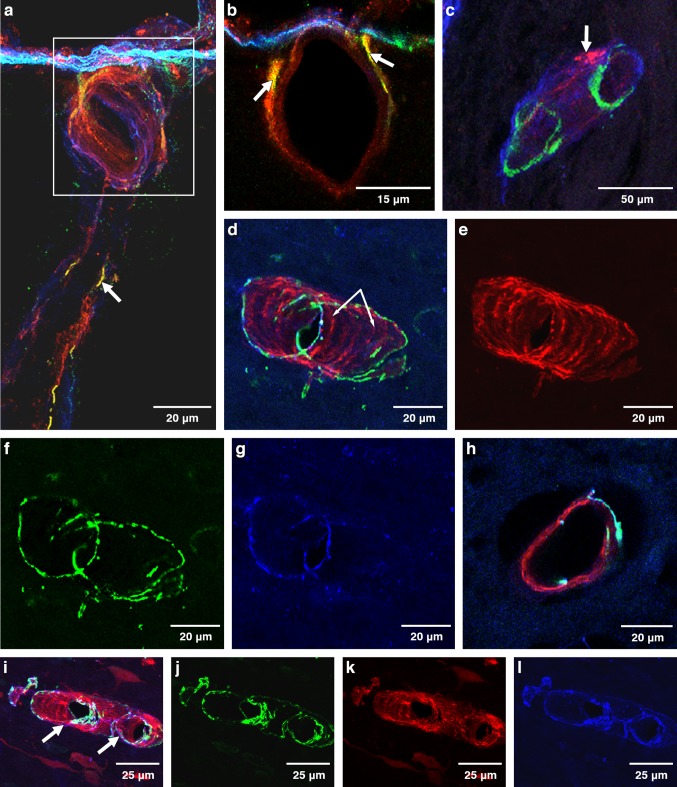
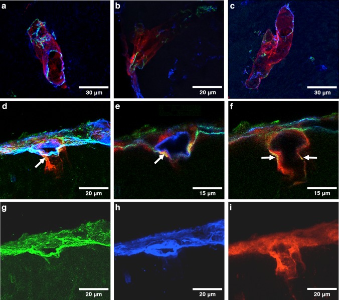
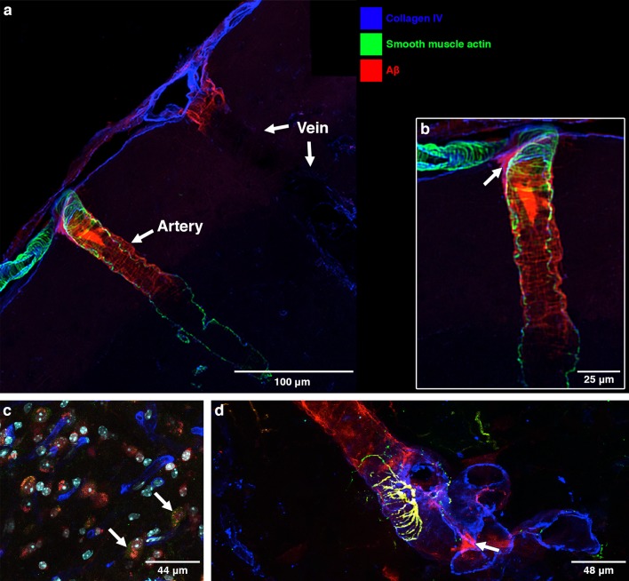
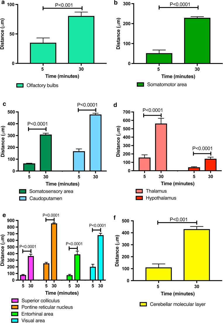
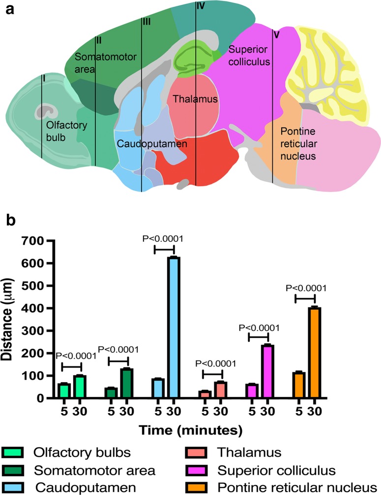
Tracers injected into CSF pass into the brain alongside arteries and out again. This has been recently termed the "glymphatic system" that proposes tracers enter the brain along periarterial "spaces" and leave the brain along the walls of veins. The object of the present study is to test the hypothesis that: (1) tracers from the CSF enter the cerebral cortex along pial-glial basement membranes as there are no perivascular "spaces" around cortical arteries, (2) tracers leave the brain along smooth muscle cell basement membranes that form the Intramural Peri-Arterial Drainage (IPAD) pathways for the elimination of interstitial fluid and solutes from the brain. 2 μL of 100 μM soluble, fluorescent fixable amyloid β (Aβ) were injected into the CSF of the cisterna magna of 6-10 and 24-30 month-old male mice and their brains were examined 5 and 30 min later. At 5 min, immunocytochemistry and confocal microscopy revealed Aβ on the outer aspects of cortical arteries colocalized with α-2 laminin in the pial-glial basement membranes. At 30 min, Aβ was colocalised with collagen IV in smooth muscle cell basement membranes in the walls of cortical arteries corresponding to the IPAD pathways. No evidence for drainage along the walls of veins was found. Measurements of the depth of penetration of tracer were taken from 11 regions of the brain. Maximum depths of penetration of tracer into the brain were achieved in the pons and caudoputamen. Conclusions drawn from the present study are that tracers injected into the CSF enter and leave the brain along separate periarterial basement membrane pathways. The exit route is along IPAD pathways in which Aβ accumulates in cerebral amyloid angiopathy (CAA) in Alzheimer's disease. Results from this study suggest that CSF may be a suitable route for delivery of therapies for neurological diseases, including CAA.

Keywords: Basement membranes; Cerebrospinal fluid; Glymphatic; Interstitial fluid; Intramural periarterial drainage.


Figures

Fig. 1
Fig. 1
Anatomical entry route of Aβ into the young adult brain from the CSF at 5 and 30 min post-injection. Young 6–10-week-old mice: Entry of amyloid-β (Aβ) tracer into the brain from the CSF at 5 min (ac). Drainage of Aβ tracer from the brain along the walls of arteries at 30 min (dl). a 5 min: the surface of the cerebral cortex (blue line) and a cortical artery passing into the brain with Aβ tracer red in its walls. Aβ tracer colocalized with α2-laminin in the pial-glial basement membrane (yellow) indicated by the white arrow. b 5 min: a single optical section of the artery enclosed in the box in a showing α2-laminin in the astrocyte basement membrane of the glia limitans on the surface of the brain (green) and co-localisation of Aβ and α2-laminin in the pial-glial basement membrane on the outer aspect of the wall of the artery (yellow) (white arrow); c 5 min: the profile of a cortical artery with smooth muscle cells (green) in its wall shows Aβ tracer (red) colocalized (pink) with collagen IV (blue) in the pial basement membrane on the outer surface of the artery (white arrows). At 30 min after injection as shown in figures (dl), the distribution of the Aβ in the artery walls is very different from that seen at 5 min in figures (ac). d An artery identified by green smooth muscle actin in its wall (f, j) shows Aβ tracer (red, e, k) colocalized (pink) with collagen IV (blue, g, l) within the wall of the artery in a spiral or ladder-type distribution (arrows in d), closely resembling the pattern of deposition of Aβ in the walls of arteries as cerebral amyloid angiopathy (CAA); h a single optical section from the artery in d) showing the Aβ tracer (red) between the smooth muscle actin staining (green)
Fig. 2
Fig. 2
Anatomical entry route of Aβ into old 24–30-month-old mouse brain from the CSF at 5 min post injection: ac arteries showing Aβ (red) along the walls of arterioles of 10–20 µm diameter labelled with collagen IV (blue) and smooth muscle actin (green); d an artery entering the cerebral cortex (arrow) showing Aβ (red) extending into the brain; e enlargement of the artery in (d) showing laminin α-2 staining in the glia limitans on the surface of the brain (green) and co-localisation of the amyloid with laminin α-2 in the vessel wall (yellow) as indicated by the arrow; f single optical section of artery in e; g single channel image of laminin α-2 from d; h single channel image of collagen IV from d; i single channel image of Aβ from d
Fig. 3
Fig. 3
Anatomical route for drainage of Aβ out of the brain following entry from the CSF at 30 min post injection in old 24–30-month-old mice: a An artery and vein at the surface of the brain. The artery is identified by smooth muscle cells in its wall (green). Aβ tracer (red) is present in a ladder-like pattern in the wall of the artery. Aβ (red) in the wall of a vein is only seen at the surface suggesting that the Aβ here has entered from the CSF and not drained from the brain. Only occasional veins had Aβ in their walls. b Enlargement of the artery in a showing the ladder-like distribution of Aβ that resembles the distribution of amyloid in CAA [29]. At the surface of the brain is a branch of a leptomeningeal artery showing the ladder-like distribution of smooth muscle cells in the tunica media. The Aβ is in the intramural periarterial drainage (IPAD) pathway; c macrophages take up Aβ tracer at 30 min after injection. The arrows indicate Aβ (red) within macrophages stained for the macrophage marker CD163 (green); d astrocytes are stained for GFAP (green). Co-localisation (yellow) of Aβ and GFAP indicates uptake of Aβ tracer by perivascular astrocytes
Fig. 4
Fig. 4
Periarterial penetration of Aβ is of greater distance after 30 min than after 5 min in cortical, subcortical and posterior brain regions of young mice. Bar charts of periarterial Aβ distance against time after injection into cisternal CSF (5 and 30 min) in the olfactory bulbs (a), somatomotor area (b), somatosensory area and caudoputamen (c), thalamus and hypothalamus (d), superior colliculus, pontine reticular nucleus, entorhinal and visual areas (e) and cerebellar molecular layer (f). Values are presented as mean ± SEM of untransformed data, with p values indicated for log10-transformed data (one-way ANOVA with Sidak’s post hoc)
Fig. 5
Fig. 5
Regional differences in periarterial distance of penetration of Aβ at 5- and 30-min post-injection in old mice. a Schematic showing a sagittal mouse brain section with five brain levels (I–V) from which distance measurements were taken in six brain regions at 5 min after injection of Aβ into cisterna magna. (Allen Institute for Brain Science. Allen Mouse Brain Atlas. Available from: http://mouse.brain-map.org/static/atlas.) b Bar charts of periarterial Aβ distance against time after injection into cisternal CSF (5 and 30 min) in the olfactory bulbs (teal), somatomotor area (green), caudoputamen (blue), thalamus (salmon), superior colliculus (pink) and pontine reticular nucleus (orange). Values are presented as mean ± SEM of untransformed data, with p values indicated for log10-transformed data (one-way ANOVA with Sidak’s post hoc)
Fig. 6
Fig. 6
Pathways for influx of CSF into the brain and drainage of CSF/ISF out of the brain along capillary and periarterial basement membranes. (1) Entry of tracers from the CSF into the brain along pial-glial basement membranes on the outer aspects of artery walls. The sites of entry of tracer into the brain extracellular compartment are unclear and could be multiple. (2) CSF enters the brain parenchyma and (3) mixes with interstitial fluid (ISF). (4) The mixture of CSF/ISF diffuses through the narrow extracellular spaces of the brain to enter (5) basement membranes in the walls of capillaries to drain out of the brain along (6) basement membranes of smooth muscle cells in the tunica media in the walls of arterioles and arteries (IPAD pathway). Key to the microanatomy of the capillary and artery walls: BM1 endothelial basement membrane. BM2 smooth muscle cell basement membrane identified in this study by the presence of collagen IV. BM3 basement membrane between pia mater (pink) and smooth muscle cell. BM4 pia-glial basement membrane between the pia mater and the astrocytes of the glia limitans. In this study, BM4 is identified by the presence of α2-laminin [9, 13]. Proposed route of IPAD tracer is located in the capillary endothelial basement membrane (5) apparently entering from the brain parenchyma. In the artery wall, tracer is observed in the basement membranes between smooth muscle cells (light purple) but not in the basement membranes on the outer aspect of the artery wall or in the endothelial basement membrane of the artery wall (both dull blue) [4, 27]. The light green arrow indicates the proposed intramural peri-arterial drainage (IPAD) pathway for fluid and solutes out of the brain

References

    1. Abbott NJ, Pizzo ME, Preston JE, Janigro D, Thorne RG. The role of brain barriers in fluid movement in the CNS: is there a ‘glymphatic’ system? Acta Neuropathol. 2018;135:387–407. doi: 10.1007/s00401-018-1812-4. - DOI - PubMed
    1. Arbel-Ornath M, Hudry E, Eikermann-Haerter K, Hou S, Gregory JL, Zhao L, Betensky RA, Frosch MP, Greenberg SM, Bacskai BJ. Interstitial fluid drainage is impaired in ischemic stroke and Alzheimer’s disease mouse models. Acta Neuropathol. 2013 - PMC - PubMed
    1. Aspelund A, Antila S, Proulx ST, Karlsen TV, Karaman S, Detmar M, Wiig H, Alitalo K. A dural lymphatic vascular system that drains brain interstitial fluid and macromolecules. J Exp Med. 2015;212:991–999. doi: 10.1084/jem.20142290. - DOI - PMC - PubMed
    1. Carare RO, Bernardes-Silva M, Newman TA, Page AM, Nicoll JA, Perry VH, Weller RO. Solutes, but not cells, drain from the brain parenchyma along basement membranes of capillaries and arteries: significance for cerebral amyloid angiopathy and neuroimmunology. Neuropathol Appl Neurobiol. 2008;34:131–144. doi: 10.1111/j.1365-2990.2007.00926.x. - DOI - PubMed
    1. Carare RO, Hawkes CA, Jeffrey M, Kalaria RN, Weller RO. Review: cerebral amyloid angiopathy, prion angiopathy, CADASIL and the spectrum of protein elimination failure angiopathies (PEFA) in neurodegenerative disease with a focus on therapy. Neuropathol Appl Neurobiol. 2013;39:593–611. doi: 10.1111/nan.12042. - DOI - PubMed

MeSH terms

LinkOut - more resources