

Nanoparticle Wettability Influences Nanoparticle-Phospholipid Interactions
- PMID: 29754486
- PMCID: PMC6507429
- DOI: 10.1021/acs.langmuir.7b03741
Nanoparticle Wettability Influences Nanoparticle-Phospholipid Interactions
Abstract
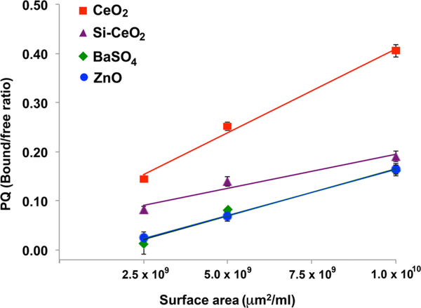
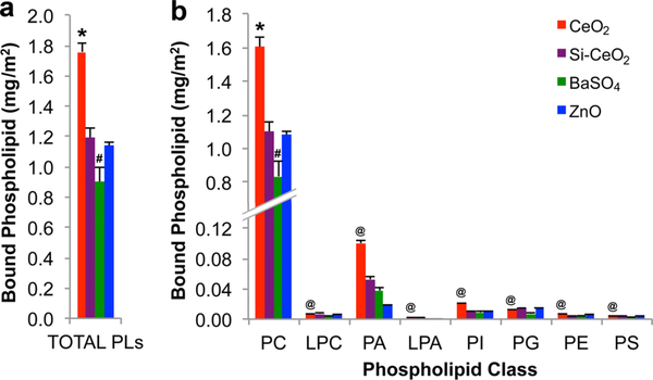
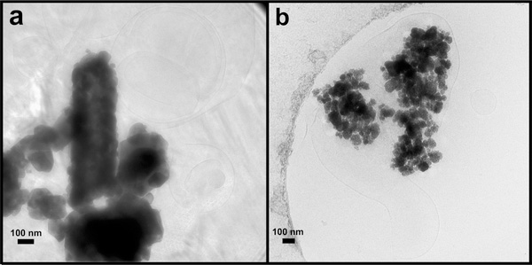
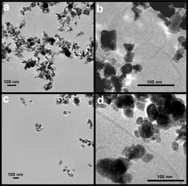
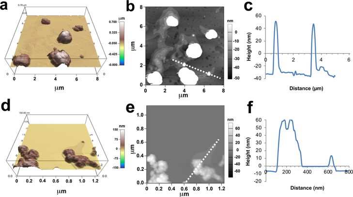
We explored the influence of nanoparticle (NP) surface charge and hydrophobicity on NP-biomolecule interactions by measuring the composition of adsorbed phospholipids on four NPs, namely, positively charged CeO2 and ZnO and negatively charged BaSO4 and silica-coated CeO2, after exposure to bronchoalveolar lavage fluid (BALf) obtained from rats, and to a mixture of neutral dipalmitoyl phosphatidylcholine (DPPC) and negatively charged dipalmitoyl phosphatidic acid (DPPA). The resulting NP-lipid interactions were examined by cryogenic transmission electron microscopy (cryo-TEM) and atomic force microscopy (AFM). Our data show that the amount of adsorbed lipids on NPs after incubation in BALf and the DPPC/DPPA mixture was higher in CeO2 than in the other NPs, qualitatively consistent with their relative hydrophobicity. The relative concentrations of specific adsorbed phospholipids on NP surfaces were different from their relative concentrations in the BALf. Sphingomyelin was not detected in the extracted lipids from the NPs despite its >20% concentration in the BALf. AFM showed that the more hydrophobic CeO2 NPs tended to be located inside lipid vesicles, whereas less hydrophobic BaSO4 NPs appeared to be outside. In addition, cryo-TEM analysis showed that CeO2 NPs were associated with the formation of multilamellar lipid bilayers, whereas BaSO4 NPs with unilamellar lipid bilayers. These data suggest that the NP surface hydrophobicity predominantly controls the amounts and types of lipids adsorbed, as well as the nature of their interaction with phospholipids.
Conflict of interest statement
The authors declare no competing financial interest.
Figures
References
-
- Monopoli MP; Åberg C; Salvati A; Dawson KA Biomolecular coronas provide the biological identity of nanosized materials. Nat. Nanotechnol 2012, 7, 779–786. - PubMed
-
- Monopoli MP; Walczyk D; Campbell A; Elia G; Lynch I; Bombelli FB; Dawson KA Physical-chemical aspects of protein corona: relevance to in vitro and in vivo biological impacts of nanoparticles. J. Am. Chem. Soc 2011, 133, 2525–2534. - PubMed
-
- Brain JD; Valberg PA Deposition of aerosol in the respiratory tract. Am. Rev. Respir. Dis 1979, 120, 1325–1373. - PubMed
-
- Buzea C; Pacheco II; Robbie K Nanomaterials and nanoparticles: sources and toxicity. Biointerphases 2007, 2, MR17–MR71. - PubMed
-
- Bastacky J; Lee CY; Goerke J; Koushafar H; Yager D; Kenaga L; Speed TP; Chen Y; Clements JA Alveolar lining layer is thin and continuous: low-temperature scanning electron microscopy of rat lung. J. Appl. Physiol 1995, 79, 1615–1628. - PubMed
Publication types
MeSH terms
Substances
Grants and funding
LinkOut - more resources
Full Text Sources
Other Literature Sources
Miscellaneous
