. 2018 Jun 7;102(6):1018-1030.
doi: 10.1016/j.ajhg.2018.03.022. Epub 2018 May 10.

Mutations in PPCS, Encoding Phosphopantothenoylcysteine Synthetase, Cause Autosomal-Recessive Dilated Cardiomyopathy

Affiliations

Mutations in PPCS, Encoding Phosphopantothenoylcysteine Synthetase, Cause Autosomal-Recessive Dilated Cardiomyopathy

Arcangela Iuso et al. Am J Hum Genet. .

Abstract

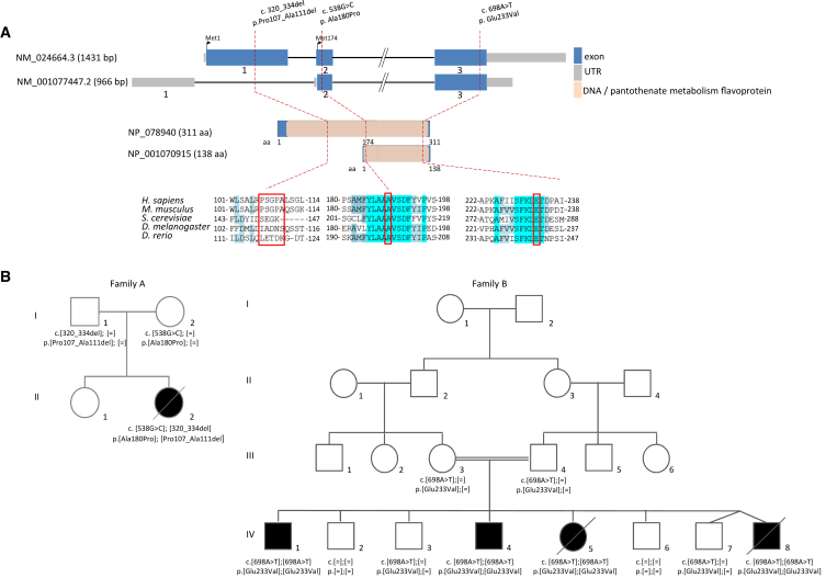
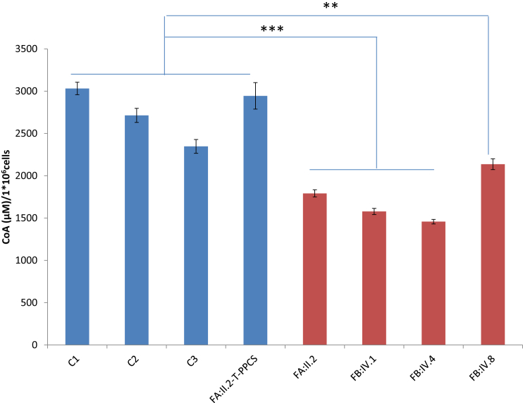
Coenzyme A (CoA) is an essential metabolic cofactor used by around 4% of cellular enzymes. Its role is to carry and transfer acetyl and acyl groups to other molecules. Cells can synthesize CoA de novo from vitamin B5 (pantothenate) through five consecutive enzymatic steps. Phosphopantothenoylcysteine synthetase (PPCS) catalyzes the second step of the pathway during which phosphopantothenate reacts with ATP and cysteine to form phosphopantothenoylcysteine. Inborn errors of CoA biosynthesis have been implicated in neurodegeneration with brain iron accumulation (NBIA), a group of rare neurological disorders characterized by accumulation of iron in the basal ganglia and progressive neurodegeneration. Exome sequencing in five individuals from two unrelated families presenting with dilated cardiomyopathy revealed biallelic mutations in PPCS, linking CoA synthesis with a cardiac phenotype. Studies in yeast and fruit flies confirmed the pathogenicity of identified mutations. Biochemical analysis revealed a decrease in CoA levels in fibroblasts of all affected individuals. CoA biosynthesis can occur with pantethine as a source independent from PPCS, suggesting pantethine as targeted treatment for the affected individuals still alive.

Keywords: PPCS; coenzyme A; dilated cardiomyopathy; pantethine treatment; pentothenate; phospohopantothenoylcysteine synthetase.


Figures

Figure 1
Figure 1
Universal Pathway for the Biosynthesis of Coenzyme A and Associated Diseases In green are indicated genes encoding either cytosolic or mitochondrial isoforms. The mitochondrial isoforms are associated with neurodegeneration with brain iron accumulation (#234200, #615643). In gray are labeled genes predicted to have mostly a cytosolic localization (Reactome: PPCS). The asterisk () indicates that the association with a disease was not reported elsewhere.
Figure 2
Figure 2
Structure of PPCS and Pedigrees of Investigated Families (A) Structure of PPCS with known conserved protein domain in the gene product and localization and conservation of amino acid residues affected by mutations identified in the two families. Intronic regions are not drawn to scale. Coloring in the sequence alignment represents the identity of amino acid residues. (B) Pedigrees of two families with mutations in PPCS. Mutation status of affected (closed symbols) and healthy (open symbols) family members. n.d., not determined.
Figure 3
Figure 3
Cardiac Magnetic Resonance Imaging (cMRI) Findings in Individuals with PPCS-Related Dilated Cardiomyopathy (A) Four-chamber view of individual IV.1 (FB:IV.1 in the figure) at the age of 21 years in diastole (Ai) and systole (Aii). Note the decreased change in ventricular volume between phases. RA, right atrium; LA, left atrium; RV, right ventricle; LV, left ventricle. (B) Short axis oblique of individual IV.4 (at the age of 10 years) in diastole (Bi) and systole (Bii), consistent with a low cardiac output. (C) Volume rendering of gadolinium-enhanced angiography of the chest and head blood vessels in individual IV.4 (posterior view), demonstrating normal vasculature.
Figure 4
Figure 4
Functional Complementation of a Yeast cab2 Deletion Mutation by Human PPCS Gene Variants Strain MGY9, used as a recipient for transformation, contains a chromosomal cab2 null mutation which was complemented by the single-copy URA3 CAB2 plasmid pGE11. Single-copy LEU2 plasmids used for complementation studies contain yeast CAB2 (positive control) and human PPCS alleles (wild-type and variants c.320_334del, c.538G>C, c.698A>T). Transformants containing two autonomously replicating plasmids are shown (left, SCD-Ura-Leu. incubation for 2 days). To counter-select URA3 plasmid pGE11, transformants were transferred to a medium supplemented with 5-FOA and incubated for 2 or 4 days (middle, right).
Figure 5
Figure 5
Western Blot Analysis of PPCS Protein in PPCS Mutant Fibroblasts (A) Western blot analysis of fibroblasts from the four affected individuals (FA:II.2, FB:IV.8, FB:IV.4, FB:IV.1), one healthy control subject (C1), and individual II.2, family A complemented by lentiviral transduction with wild-type PPCS. 7.5 and 15 μg proteins were loaded. Tubulin was used as a loading control. (B) Densitometric analysis of this western blot. Error bars indicate the standard error of the mean. (C) Blue native gel electrophoresis of total cellular extract from individual II.2, family A, and control C1 solubilized by 1% laurylmaltoside. ATP5α, sub-unit of the respiratory chain complex V, was used as a loading control
Figure 6
Figure 6
Total Cellular CoA in Fibroblasts Fibroblasts growing in exponential phase under standard medium were collected, homogenized, and evaluated for CoA content. Relative fluorescence values (RFU) of serial dilution of CoA standard were measured to generate a calibration curve. CoA levels in fibroblasts were calculated by making use of the calibration curve. Fibroblasts from three unrelated controls (C1, C2, C3 in the figure) and from an affected individual transduced with wild-type PPCS (FA:II.2-T-PPCS) are labeled in blue; affected individuals (FA:II.2, FB:IV.1, FB:IV.4, FB:IV.8) are labeled in red. Results are mean ± SD of n = 16 values. p values were calculated with an independent sample t test. All p values were two-sided with a significance level of 0.05.
Figure 7
Figure 7
Physiological Cardiac Parameters Are Changed in Homozygous dPPCS1Drosophila melanogaster Parameters: (A) heart rate in beats per minute, (B) arrhythmia index, (C) systolic and (D) diastolic length in seconds, (E) heart wall shortening, and (F) representative kymographs (10 s). ∗∗p ≤ 0.01 and ∗∗∗p ≤ 0.001. N = 50 for heterozygous and N = 36 for homozygous for (A) and (B); N = 18 for heterozygous and N = 15 for homozygous for (C), (D), and (E).

References

    1. Hartig M.B., Hörtnagel K., Garavaglia B., Zorzi G., Kmiec T., Klopstock T., Rostasy K., Svetel M., Kostic V.S., Schuelke M. Genotypic and phenotypic spectrum of PANK2 mutations in patients with neurodegeneration with brain iron accumulation. Ann. Neurol. 2006;59:248–256. - PubMed
    1. Dusi S., Valletta L., Haack T.B., Tsuchiya Y., Venco P., Pasqualato S., Goffrini P., Tigano M., Demchenko N., Wieland T. Exome sequence reveals mutations in CoA synthase as a cause of neurodegeneration with brain iron accumulation. Am. J. Hum. Genet. 2014;94:11–22. - PMC - PubMed
    1. Hogarth P. Neurodegeneration with brain iron accumulation: diagnosis and management. J. Mov. Disord. 2015;8:1–13. - PMC - PubMed
    1. Annesi G., Gagliardi M., Iannello G., Quattrone A., Iannello G., Quattrone A. Mutational analysis of COASY in an Italian patient with NBIA. Parkinsonism Relat. Disord. 2016;28:150–151. - PubMed
    1. Evers C., Seitz A., Assmann B., Opladen T., Karch S., Hinderhofer K., Granzow M., Paramasivam N., Eils R., Diessl N. Diagnosis of CoPAN by whole exome sequencing: Waking up a sleeping tiger’s eye. Am. J. Med. Genet. A. 2017;173:1878–1886. - PubMed

Publication types

MeSH terms