. 2018 May 21;45(4):512-525.e5.
doi: 10.1016/j.devcel.2018.04.013. Epub 2018 May 10.

Intracellular Calcium Mobilization Is Required for Sonic Hedgehog Signaling

Affiliations

Intracellular Calcium Mobilization Is Required for Sonic Hedgehog Signaling

Dana Klatt Shaw et al. Dev Cell. .

Abstract

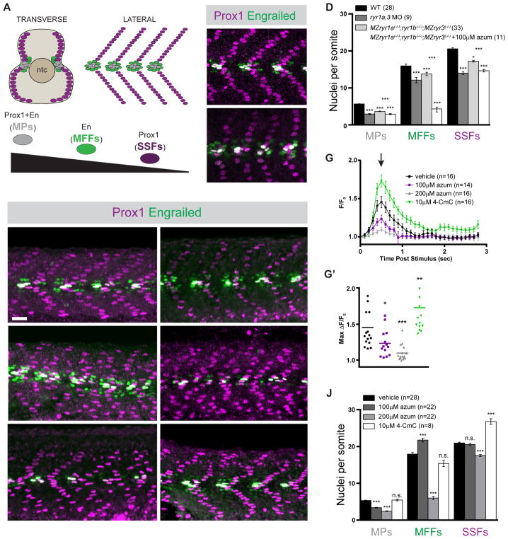
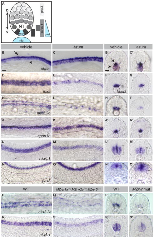
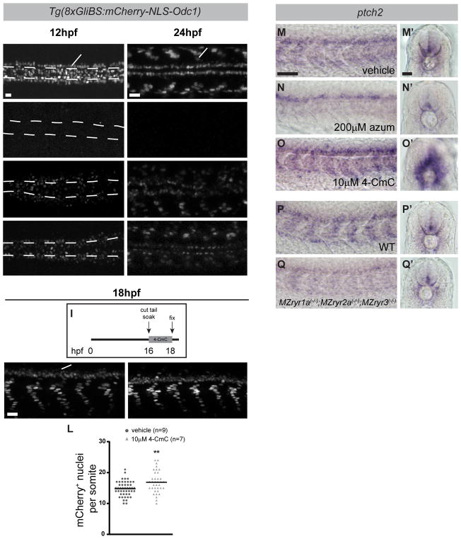
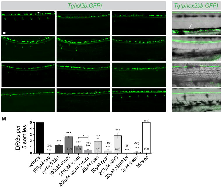
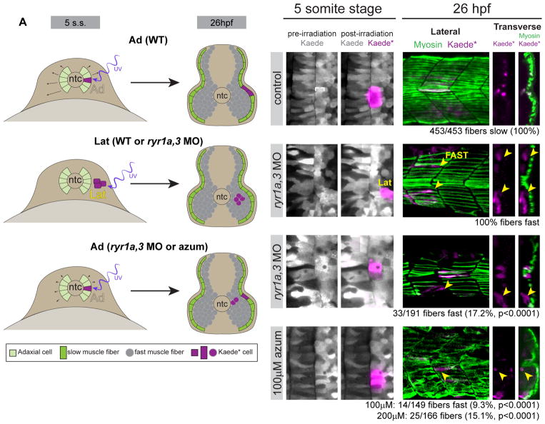
Graded Shh signaling across fields of precursor cells coordinates patterns of gene expression, differentiation, and morphogenetic behavior as precursors form complex structures, such as the nervous system, the limbs, and craniofacial skeleton. Here we discover that intracellular calcium mobilization, a process tightly controlled and readily modulated, regulates the level of Shh-dependent gene expression in responding cells and affects the development of all Shh-dependent cell types in the zebrafish embryo. Reduced expression or modified activity of ryanodine receptor (RyR) intracellular calcium release channels shifted the allocation of Shh-dependent cell fates in the somitic muscle and neural tube. Mosaic analysis revealed that RyR-mediated calcium mobilization is required specifically in Shh ligand-receiving cells. This work reveals that RyR channels participate in intercellular signal transduction events. As modulation of RyR activity modifies tissue patterning, we hypothesize that alterations in intracellular calcium mobilization contribute to both birth defects and evolutionary modifications of morphology.

Keywords: hedgehog signaling; intracellular calcium release; ryanodine receptors; tissue pattering.


Conflict of interest statement

COMPETING FINANCIAL INTERESTS

The authors declare no competing financial interests.

Figures

Figure 1
Figure 1
Formation of Shh-dependent muscle requires RyR function. (A) Schematic illustration of the nuclei of Shh-dependent muscle cells in the somites of 26hpf zebrafish embryos, presented relative to the notochord (ntc) in transverse section and in a superficial lateral view (anterior to left). Nuclei of different muscle types are color-coded: Slow Muscle Pioneer Cells (MPs, grey), Medial Fast Fibers (MFFs, green), and Superficial Slow Fibers (SSFs, magenta). (B, C, E, F, H, I, K, L) Shh-dependent muscle fiber nuclei, marked by Prox1 (magenta) and En (green) expression, were visualized by immunostaining 26hpf embryos: (B) wildtype (WT), (C) ryr1a and ryr3 MO-injected (ryr1a,3 MO), (E) vehicle-treated (0.5% DMSO) WT, (F) MZryr1a(−/−);ryr1b(−/−);MZryr3(−/−) mutant, (H and I) azumolene-treated WT, (K) azumolene-treated MZryr1a(−/−);ryr1b(−/−);MZryr3(−/−) mutant, and (L) 4-CmC-treated WT (supernumerary Prox1+ SSF nuclei are indicated with arrowheads). All nuclei co-expressing En and Prox1 were considered MPs. Scale bar indicates 25 μm. (D and J) Quantification of distinct muscle cell types per somite. Data are represented as mean ±SEM. As numbers of nuclei in WT and DMSO-treated embryos did not differ significantly, comparisons were made to control vehicle-treated embryos unless otherwise indicated. (G) Traces of GCaMP fluorescence in electrically stimulated muscle of 24hpf Tg(act2b:GCaMP6f) embryos incubated from 6 to 24hpf in indicated solutions. Data are represented as mean ±SEM. The maximum change in fluorescence from baseline (arrow in G) is shown for each recorded embryo in (G′) with horizontal lines representing means. See also Figure S2.
Figure 2
Figure 2
RyR function is required for Shh-dependent patterning of the neural tube. (A) Schematic of Shh-dependent neural tube (NT) patterning. The medial floorplate (MFP), lateral floorplate (LFP), and neighboring motoneurons (mn) are indicated. The notochord (ntc) and MFP are sources of Shh, designated in blue. Approximate gene expression domains of nkx2.2b, isl1, nkx6.1, and pax3 are indicated. (B–O) Whole mount in situ hybridization to detect neural tube markers in vehicle-treated (0.5% DMSO) and 200μM azumolene-treated embryos. (B,C) shha-expression in the notochord (arrowhead) and MFP (arrow) is similar in control and treated embryos. (D–O) Gene expression in the neural tube is shifted upon azumolene treatment in a manner consistent with reduced Shh signaling. (D–G) Whereas foxa and foxa2 are expressed in both the MFP and LFP in control embryos, they are expressed in fewer FP cells in azumolene-treated embryos. (H–K) Expression of nkx2.2b marking the LFP is reduced, but spon1b expression marking the MFP appears unchanged in azumolene-treated embryos. (L–O) The ventral domain of the neural tube, marked by nkx6.1, is reduced, while the dorsal domain of the neural tube, marked by pax3, is expanded in azumolene-treated embryos. (P–S) Expression of nkx2.2a in the LFP and nkx6.1 in the ventral domain of the neural tube is diminished in MZryr1a(−/−);MZryr2a(−/−);MZryr3(−/−) as compared with WT. B,C,L,M,N,O,R,S are lateral views of flat mounted embryos. D,E,H,I,J,K,P,Q are dorsal views of flat mounted embryos. Prime letters, F, and G are transverse cross sections. Scale bars indicate 25μm. Interval bars indicate dorsoventral extents of gene expression domains. See also Figure S3.
Figure 3
Figure 3
RyR function is required for Shh-dependent Gli-mediated gene expression. (A–H) Dorsal view images of live Tg(8xGli:mCherry-NLS-Odc1) embryos at 12hpf (A,C,E,G) or 24hpf (B,D,F,H) that had been treated with vehicle (0.5%DMSO), cyclopamine, azumolene, or ryanodine. At 12hpf, the position of the notochord (ntc) just ventral to the FP is outlined by dashed lines. (A) In control 12hpf embryos, mCherry is expressed in nuclei of cells responding to Shh, including adaxial cells (arrowhead) and FP cells (arrow). (B) At 24hpf, mCherry is expressed in nuclei of slow muscle cells (arrowhead) and cells in the ventral neural tube (arrow). (C–H) mCherry expression is reduced in embryos treated with each drug. (I) Tail-transected Tg(8xGli:mCherry-NLS-Odc1) embryos were soaked in vehicle or 4-CmC from 16 to 18hpf, fixed, and imaged. (J–L) Potentiation of RyR channel activity with 4-CmC treatment results in increased numbers of presumptive slow muscle nuclei that express the mCherry reporter (J and K are lateral views, arrowhead indicates nuclei of slow muscle cells and arrow indicates cells in the ventral neural tube). (L) Quantification of mCherry+ nuclei per somite in 4-CmC-treated embryos. Each point represents a single somite and the horizontal line represents the mean. (M–Q) RyR activity affects endogenous ptch2 expression as detected by whole mount in situ hybridization in 24hpf embryos. (M–Q) Lateral views reveal expression in somites and (M′–Q′) transverse sections reveal expression in slow muscle cells surrounding the notochord and in the ventral neural tube. As compared with WT embryos, ptch2 expression is diminished azumolene-treated and MZryr1a(−/−);MZryr2a(−/−);MZryr3(−/−) mutant embryos, and it is enhanced in 4-CmC-treated embryos. Scale bars indicate 25 μm.
Figure 4
Figure 4
RyR function is required for development of Shh-dependent neural crest-derived neurons of the dorsal root ganglia (DRGs) and the enteric nervous system (ENS). (A–L) Lateral views of live 72hpf Tg(isl2b:GFP) embryos with GFP-labeled Rohon Beard neurons (arrow) and DRGs (arrowhead). Embryos were treated between 24 and 48hpf with cyclopamine (cyc), azumolene (azum), ryanodine (ryan), N-acetyl cysteine (NAC), aldrithiol, thapsigargin (thaps), or tricaine at indicated concentrations or injected at the one-cell stage with ryr1a and ryr3 MOs. Arrowheads in C and G indicate the presence of small, faint DRGs. (M) Quantification of embryos treated as in A–L. DRGs present in somites 11–15 were counted. For each condition, the number of embryos analyzed is indicated. Comparisons are to control vehicle-treated embryos unless otherwise indicated. Data are represented as mean ±SEM. (N–Q) Lateral views of live 78hpf Tg(phox2bb:GFP) embryos with the ENS (arrow) labeled by GFP in control, cyclopamine-treated, ryr1a,3 morphant, or ryanodine-treated embryos. Black melanophores (pigment) are visible in each condition. Scale bar in A indicates 25 μm. See also Figure S4.
Figure 5
Figure 5
Adaxial precursors transfate to produce Shh-independent fast muscle derivatives in embryos with diminished RyR function. (A) Schematic representation of experimental setup and summary of results, with embryos shown in transverse views. At the 5-somite stage Kaede was photoconverted to Kaede* in small groups of adaxial (Ad) slow muscle precursors or lateral (Lat) fast muscle precursors. At 26hpf (14 hours later), embryos were fixed and stained for F59 myosin expression. (B,D,F,H) Labeling of muscle precursor cells for lineage analysis. Clusters of slow muscle precursor adaxial (B–B′, F–F′, H–H′) or fast muscle precursor lateral (D–D′) cells were labeled by photoconversion (magenta) at the 5-somite stage. At 26hpf, the positions of Kaede*-labeled (magenta) cells in the somites were determined relative to the superficial slow muscle, detected with the F59 antibody (myosin, green). (C–C″) Kaede*-labeled adaxial cells always gave rise to F59+ descendants in the parallel array of superficial slow muscle cells (white arrowheads) in control embryos. (E–E″) Kaede*-labeled lateral somite precursors always became deep fast muscle regardless of ryr gene expression (yellow arrowheads). (G–G″ and I–I″) In ryr1a,3 morphant or azumolene-treated embryos, adaxial cells gave rise to both slow (white arrowheads) and fast (yellow arrowheads) muscle fibers.
Figure 6
Figure 6
RyR function is required in the Shh ligand-receiving cell. (A) Design of mosaic analysis experiments. Spatially separated NC progenitors and dorsal organizer (shield) are indicated. Formation of donor-derived Rohon Beard (RB) and Dorsal Root Ganglia (DRG) neurons in mosaic embryos was determined at 78hpf. (B–D) Examples of chimeric embryos indicating the differentiation of donor-labeled Tg(isl2b:GFP) tissue in unlabeled hosts. (B and C) Donor wild type (WT) cells gave rise to both DRGs (arrows) and RBs (arrowheads) when they developed in either WT or ryr1a,3 morphant hosts. (D) ryr1a,3 morphant cells failed to produce DRGs in WT hosts. (E) The numbers of GFP-labeled RBs and DRGs present in each mosaic embryo is plotted. Lines representing best-fit analyses indicate that WT host donor cells gave rise to DRGs and RBs in a 2 to 3 ratio (black and magenta lines with slopes of 0.64 and 0.69, respectively). In contrast, donor cells from ryr1a,3 morphant embryos gave rise to RBs, but not DRGs (green line with a slope of 0.06). (F–M) Prox1 and En expression at 26hpf in (F and G) shha RNA-injected, (H and I) MZptch2(−/−), (J and K) SmoM2 RNA-injected, and (L and M) dnPKA RNA-injected embryos. Azumolene treatment attenuated overproduction of Shh-dependent muscle cell types activated by Shh overexpression, loss of the Patched2 receptor, or overexpression of SmoM2. In contrast, azumolene failed to alter the development of muscle cells in embryos expressing dnPKA. Scale bar in F indicates 25 μm. See also Figures S5 and S6.

Similar articles

Cited by

References

    1. Abzhanov A, Kuo WP, Hartmann C, Grant BR, Grant PR, Tabin CJ. The calmodulin pathway and evolution of elongated beak morphology in Darwin’s finches. Nature. 2006;442:563–567. - PubMed
    1. Attili S, Hughes SM. Anaesthetic tricaine acts preferentially on neural voltage-gated sodium channels and fails to block directly evoked muscle contraction. PLoS One. 2014;9:e103751. - PMC - PubMed
    1. Barresi MJ, Stickney HL, Devoto SH. The zebrafish slow-muscle-omitted gene product is required for Hedgehog signal transduction and the development of slow muscle identity. Development. 2000;127:2189–2199. - PubMed
    1. Beachy PA, Karhadkar SS, Berman DM. Tissue repair and stem cell renewal in carcinogenesis. Nature. 2004;432:324–331. - PubMed
    1. Belgacem YH, Borodinsky LN. Sonic hedgehog signaling is decoded by calcium spike activity in the developing spinal cord. Proc Natl Acad Sci U S A. 2011;108:4482–4487. - PMC - PubMed

Publication types

MeSH terms