. 2018 Jul 3;26(7):925-935.e8.
doi: 10.1016/j.str.2018.04.003. Epub 2018 May 10.

Structure of Human NatA and Its Regulation by the Huntingtin Interacting Protein HYPK

Affiliations

Structure of Human NatA and Its Regulation by the Huntingtin Interacting Protein HYPK

Leah Gottlieb et al. Structure. .

Abstract

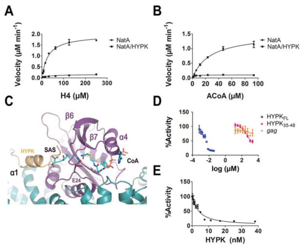
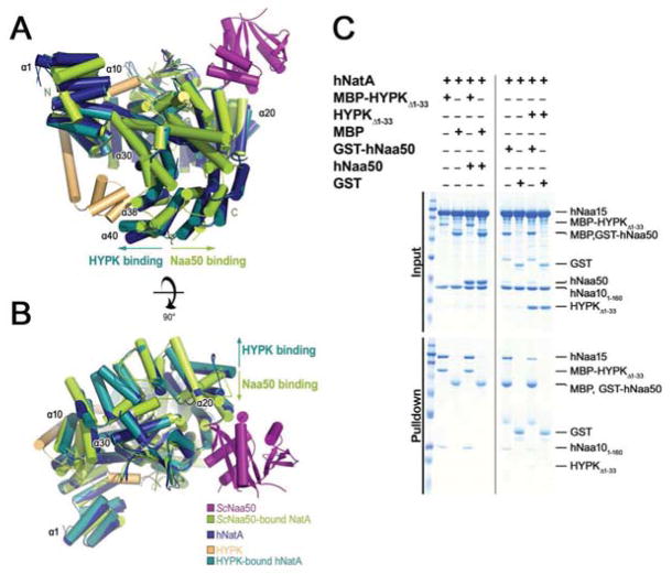
Co-translational N-terminal protein acetylation regulates many protein functions including degradation, folding, interprotein interactions, and targeting. Human NatA (hNatA), one of six conserved metazoan N-terminal acetyltransferases, contains Naa10 catalytic and Naa15 auxiliary subunits, and associates with the intrinsically disordered Huntingtin yeast two-hybrid protein K (HYPK). We report on the crystal structures of hNatA and hNatA/HYPK, and associated biochemical and enzymatic analyses. We demonstrate that hNatA contains unique features: a stabilizing inositol hexaphosphate (IP6) molecule and a metazoan-specific Naa15 domain that mediates high-affinity HYPK binding. We find that HYPK harbors intrinsic hNatA-specific inhibitory activity through a bipartite structure: a ubiquitin-associated domain that binds a hNaa15 metazoan-specific region and an N-terminal loop-helix region that distorts the hNaa10 active site. We show that HYPK binding blocks hNaa50 targeting to hNatA, likely limiting Naa50 ribosome localization in vivo. These studies provide a model for metazoan NAT activity and HYPK regulation of N-terminal acetylation.

Keywords: HYPK; Huntington interacting protein; N-terminal acetylation; NatA; X-ray crystallography; protein complex.


Conflict of interest statement

Declaration of Interests

The authors declare no competing interests.

Figures

Figure 1
Figure 1. Overall Structure of Human NatA
(A) hNaa10 (light blue) and hNaa15 (dark blue) shown in cartoon overlayed with S. pombe (Sp) NatA (dark grey) (pdb: 4KVO). Electron density for α33 and α34 helices, annotated with an asterisk (*), was not resolved in the human structure. (B) A 90° view rotation of A. with bound inositol hexaphosphate (IP6) in stick format and annotated by a dashed box. (C) Zoom view depicting key residues involved in interactions with IP6. (D) Zoom view of the hydrophobic pocket formed at the interface of the hNaa15 α22–α23 helices and C-terminal α44–45 helices. (E) Sedimentation velocity of hNatA mutants of Y834 (yellow in D). WT, wild-type. (F) Differential scanning calorimetric analysis of hNatA mutants. (G) Bar graph showing the initial velocities of mutants (Py834F=0.0001 and PY834A<0.0001 by Student’s unpaired, two-tailed t-test). Assays performed in triplicate; error bars S.E.M.
Figure 2
Figure 2. Specific Binding and Inhibition by HYPK to Human NatA
(A) Bar graph showing the relative effect of HYPK presence on Nt- (NAT) and Histone acetyltransferase (HAT) activity (PhNatA=0.0003, PhPCAF=0.0148, and PhNatF=0.0253, by Student’s unpaired, two-tailed t-test). Reactions were performed in triplicate; error bars S.E.M. (B) MBP pull-down assay comparing the ability of HYPK to bind to the human and SpNatA complexes.
Figure 3
Figure 3. Structure of HYPK-Bound Human NatA
(A) Sequence alignment of HYPK with Nascent polypeptide-Associated Complex (NAC) proteins from M. thermautotrophicus (Mt); S. cerevisiae (Sc); H. sapiens (Hs); and S. pombe (Sp). Secondary structure determined from hNatA/HYPK structure shown with hNaa10 (black square) and hNaa15 contacts (red circle) in the middle. Dashed line indicates construct used for crystallization. Ubiquitin-Associated domain (UBA) indicated by black bar. (B) HYPK-bound hNaa10 (purple) and hNaa15 (dark teal) shown bound to HYPK (light orange) in cartoon with bound IP6 (stick format). (C) HYPK-bound hNatA complex shown in cartoon superimposed with hNaa10 (light cyan) and hNaa15 (dark blue). hNaa10 α4 is annotated in yellow. (D) A 90° view rotation of C. (E) Zoom view of the interface between the HYPK UBA domain (90–129) and the hNaa15 α38 and α40 helices. (F) Zoom view of HYPK-bound hNaa15 in E. in tube and stick format depicting residues involved in HYPK-hNaa15 interaction. (G) A 180° view rotation of F. (H) Zoom view of the hNaa10 active site demonstrating the obstruction and rearrangement of key active site residues by HYPK. (I) Zoom view of Y49. (J) Zoom view of E74.
Figure 4
Figure 4. Molecular and Thermodynamic Details of HYPK-hNatA Interaction
(A) Differential scanning calorimetric analysis comparing hNatA and hNatA/HYPK complexes. (B) MBP pull-down assay evaluating the effects of mutations on critical HYPK residues or HYPK truncation on the hNatA binding. (C) Bar graph evaluating the effect of mutant and truncation HYPK constructs on hNatA inhibition potency. Assay performed in triplicate; error bars S.E.M. WT, wild type.
Figure 5
Figure 5. Mechanism of NatA Inhibition
Michaelis-Menten kinetics of hNatA and hNatA/HYPK complexes (10 nM) with respect to (A) H4 (B) Acetyl-CoA (C) Dose-response curve corresponding to titration of full-length MBP-HYPK recombinant protein, HYPK peptide (containing the residues 35–48), and the non-specific gag peptide. (D) Overlay of the SASE-CoA bisubstrate inhibitor (pdb: 4KVM, CMK stick format) bound in the hNatA-HYPK active site. (E) Morrison inhibition plot of the dose-response curve corresponding solely to the titration of full-length MBP-HYPK recombinant protein. Assays performed in triplicate; error bars correspond to the S.E.M. for each point.
Figure 6
Figure 6. HYPK Binding Reduces Human NatA Capacity to Bind Naa50
(A) Overlay of S. cerevisiae (Sc) Naa50-bound NatA (pdb: 4XPD; ScNatA: green; ScNaa50: magenta) with hNatA (dark blue) and HYPK (light orange) bound to hNatA (dark teal). Helices are illustrated in cylindrical cartoons and enumeration of human NatA helices are indicated. Arrows indicate the direction of Naa15 conformational change induced upon HYPK (dark teal) or Naa50 (green) binding. (B) A 90° view rotation of A. (C) Competition pull-downs of HYPK and hNaa50 binding to hNatA with MBP-HYPKΔ1–33 as bait in the presence of amylose resin (left) and GST-hNaa50 as bait in the presence of glutathione resin (right).

References

    1. Adams PD, Afonine PV, Bunkoczi G, Chen VB, Davis IW, Echols N, Headd JJ, Hung LW, Kapral GJ, Grosse-Kunstleve RW, et al. PHENIX: a comprehensive Python-based system for macromolecular structure solution. Acta Crystallogr D Biol Crystallogr. 2010;66:213–221. - PMC - PubMed
    1. Aksnes H, Drazic A, Marie M, Arnesen T. First Things First: Vital Protein Marks by N-Terminal Acetyltransferases. Trends Biochem Sci. 2016;41:746–760. - PubMed
    1. Aksnes H, Van Damme P, Goris M, Starheim KK, Marie M, Støve SI, Hoel C, Kalvik TV, Hole K, Glomnes N, et al. An organellar nα-acetyltransferase, naa60, acetylates cytosolic N termini of transmembrane proteins and maintains Golgi integrity. Cell Reports. 2015;10:1362–1374. - PubMed
    1. Andersen KM, Semple CA, Hartmann-Petersen R. Characterisation of the nascent polypeptide-associated complex in fission yeast. Mol Biol Rep. 2007;34:275–281. - PubMed
    1. Arnesen T, Starheim KK, Van Damme P, Evjenth R, Dinh H, Betts MJ, Ryningen A, Vandekerckhove J, Gevaert K, Anderson D. The chaperone-like protein HYPK acts together with NatA in cotranslational N-terminal acetylation and prevention of Huntingtin aggregation. Mol Cell Biol. 2010;30:1898–1909. - PMC - PubMed

Publication types

LinkOut - more resources