. 2018 Apr 27:9:903.
doi: 10.3389/fimmu.2018.00903. eCollection 2018.

Differential Effect of Viable Versus Necrotic Neutrophils on Mycobacterium tuberculosis Growth and Cytokine Induction in Whole Blood

Affiliations

Differential Effect of Viable Versus Necrotic Neutrophils on Mycobacterium tuberculosis Growth and Cytokine Induction in Whole Blood

David M Lowe et al. Front Immunol. .

Abstract

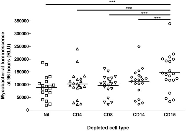
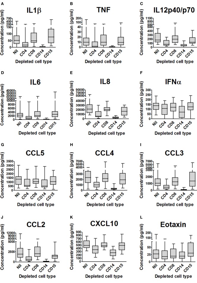
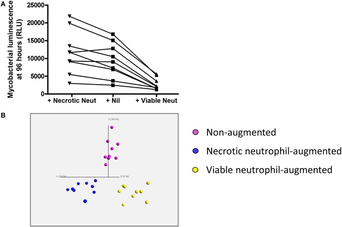
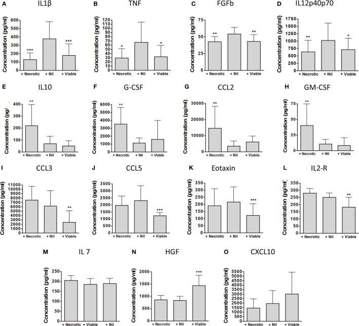
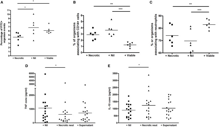
Neutrophils exert both positive and negative influences on the host response to tuberculosis, but the mechanisms by which these differential effects are mediated are unknown. We studied the impact of live and dead neutrophils on the control of Mycobacterium tuberculosis using a whole blood bioluminescence-based assay, and assayed supernatant cytokine concentrations using Luminex™ technology and ELISA. CD15+ granulocyte depletion from blood prior to infection with M. tuberculosis-lux impaired control of mycobacteria by 96 h, with a greater effect than depletion of CD4+, CD8+, or CD14+ cells (p < 0.001). Augmentation of blood with viable granulocytes significantly improved control of mycobacteria by 96 h (p = 0.001), but augmentation with necrotic granulocytes had the opposite effect (p = 0.01). Both augmentations decreased supernatant concentrations of tumor necrosis factor and interleukin (IL)-12 p40/p70, but necrotic granulocyte augmentation also increased concentrations of IL-10, G-CSF, GM-CSF, and CCL2. Necrotic neutrophil augmentation reduced phagocytosis of FITC-labeled M. bovis BCG by all phagocytes, whereas viable neutrophil augmentation specifically reduced early uptake by CD14+ cells. The immunosuppressive effect of dead neutrophils required necrotic debris rather than supernatant. We conclude that viable neutrophils enhance control of M. tuberculosis in blood, but necrotic neutrophils have the opposite effect-the latter associated with induction of IL-10, growth factors, and chemoattractants. Our findings suggest a mechanism by which necrotic neutrophils may exert detrimental effects on the host response in active tuberculosis.

Keywords: mycobacteria; necrosis; neutrophil; tuberculosis; viability.


Figures

Figure 1
Figure 1
Impact of cell depletions on control of mycobacterial luminescence in whole blood. Blood was taken from 20 donors. 450 mcl of blood depleted of CD4+, CD8+, CD14+, CD15+ or no cells (in triplicate per donor per condition), diluted 1:1 with RPMI-1640, was infected with approximately 5 × 105 CFU Mycobacterium tuberculosis-lux in 100 mcl PBS. Samples were incubated on a rocking plate (20 rpm) at 37°C for 96 h before removal of supernatants, lysis of red blood cells with water, resuspension in 1 ml PBS and measurement of mycobacterial luminescence on at least two 100 mcl aliquots. Results presented are the mean of all measurements across triplicate samples for the relevant condition. ***p < 0.001 (one-way ANOVA).
Figure 2
Figure 2
Impact of cell depletions on supernatant cytokines and chemokines from Mycobacterium tuberculosis-infected blood. (A–R). Supernatants from the experiments presented in Figure 1 (blood from 20 donors depleted of CD4+, CD8+, CD14+, CD15+ or no cells and infected with M. tuberculosis-lux for 96 h) were aspirated and stored at −80°C until analysis by Luminex™ technology. Box and whisker plots represent median, interquartile range, minimum and maximum values for each analyte. Abbreviations: VEGF, vascular endothelial growth factor; TNF, tumor necrosis factor; IFN, interferon; IL, interleukin; IL-2R, interleukin-2 receptor; IL-1RA, interleukin-1 receptor antagonist; FGFb, fibroblast growth factor basic; EGF, epidermal growth factor; CXCL, C-X-C motif ligand; CCL, C-C chemokine ligand; CD, cluster of differentiation.
Figure 2
Figure 2
Impact of cell depletions on supernatant cytokines and chemokines from Mycobacterium tuberculosis-infected blood. (A–R). Supernatants from the experiments presented in Figure 1 (blood from 20 donors depleted of CD4+, CD8+, CD14+, CD15+ or no cells and infected with M. tuberculosis-lux for 96 h) were aspirated and stored at −80°C until analysis by Luminex™ technology. Box and whisker plots represent median, interquartile range, minimum and maximum values for each analyte. Abbreviations: VEGF, vascular endothelial growth factor; TNF, tumor necrosis factor; IFN, interferon; IL, interleukin; IL-2R, interleukin-2 receptor; IL-1RA, interleukin-1 receptor antagonist; FGFb, fibroblast growth factor basic; EGF, epidermal growth factor; CXCL, C-X-C motif ligand; CCL, C-C chemokine ligand; CD, cluster of differentiation.
Figure 3
Figure 3
Impact of viable and necrotic neutrophil augmentation on control of mycobacterial luminescence by whole blood and on supernatant cytokines and chemokines from Mycobacterium tuberculosis-infected blood. (A) 100,000 RLU of M. tuberculosis-lux in 100 mcl PBS was inoculated into samples of 450 mcl whole blood plus either 450 mcl Percoll-isolated autologous neutrophils in RPMI-1640 heat-shocked at 60°C for 20 min and allowed to cool (“+Necrotic Neut”), 450 mcl RPMI-1640 only (“+Nil”), or 450 mcl room temperature Percoll-isolated autologous neutrophils in RPMI-1640 (“+Viable Neut”). After 96-h incubation, red blood cells were lysed and luminescence was measured on at least two aliquots of 100 mcl. Results are shown from nine independent donors. (B) Three-dimensional principal component analysis (PCA) plot generated using cytokine/chemokines that significantly contribute to differentiation between supernatants of augmentation conditions (calculated using multi-group comparison; purple = non-augmented, blue = necrotic neutrophil-augmented, yellow = viable neutrophil-augmented). PCA is a technique to reduce the dimensionality of complex datasets by transforming the data to a coordinate system. The first three coordinates (principal components) are represented as a 3D plot. The first principal component accounts for as much variability as possible within the data, and each succeeding component accounts for the next highest proportion of the variability possible, but under the constraint that it is not correlated with preceding components. This allows visualization of the differences between patient samples and analytes within complex datasets. Individual points represent one donor in one augmentation condition and their position in the plot is determined by the combined effects of all parameters measured for the sample that significantly contribute to the overall between-group difference. Component vectors for the three main components are displayed, along with a percentage figure signifying the proportion of the variability in the data that each component accounts for. Analysis is presented using raw values from infected supernatants.
Figure 4
Figure 4
Detailed impact of viable and necrotic neutrophil augmentation on supernatant cytokines and chemokines from Mycobacterium tuberculosis-infected blood. (A–O) Cytokines and chemokines were measured in 96-h supernatants of M. tuberculosis-infected blood which had been augmented with necrotic autologous neutrophils (“+Necrotic”), with medium only (“+Nil”), or with viable autologous neutrophils (“+Viable”). Bars represent mean ± SD for each analyte found to be significant in principal component analysis. Presented are p-values from one-way ANOVA with Bonferroni correction comparing both augmented conditions with the medium-only control: *p < 0.05, **p < 0.01, ***p < 0.001. Data are from nine separate donors in four independent experiments. Abbreviations: IL, interleukin; TNF, tumor necrosis factor; FGFb, fibroblast growth factor basic; G-CSF, granulocyte-colony stimulating factor; GM-CSF, granulocyte macrophage-colony stimulating factor; CCL, C-C chemokine ligand; CXCL, C-X-C motif ligand; IL-2R, interleukin-2 receptor; HGF, hepatocyte growth factor.
Figure 5
Figure 5
Effect of viable and necrotic neutrophil augmentation on phagocytosis of FITC-labeled M. bovis BCG, and differential effect of necrotic neutrophil augmentation versus supernatant of necrotic neutrophils on cytokine release in blood. (A–C) Blood from six donors (four independent experiments) was augmented with viable neutrophils, necrotic neutrophils, or medium alone, infected with 2 × 105 CFU FITC-labeled M. bovis BCG and incubated for 1 h at 37°C. 2 × 100 mcl aliquots were taken from each sample, incubated with CD14-PE and Viability Dye for 20 min, red blood cells were lysed, trypan blue was added to quench extracellular fluorescence and samples fixed in 2% paraformaldehyde before acquisition on a BD Fortessa flow cytometer. Results are presented as the percentage of BCG-FITC+ events (A), the percentage of BCG-FITC+ events associated with CD14+ cells (B), and the percentage of BCG-FITC+ events associated with neutrophils [as defined by forward and side scatter among CD14 negative events (C)]. (D,E) Blood from 17 donors (4 independent experiments) was augmented with necrotic neutrophils, the supernatant of necrotic neutrophils, or medium alone, infected with 2 × 105 CFU M. bovis BCG and incubated for 96 h at 37°C. Samples were centrifuged and supernatants stored at −80°C until analysis by ELISA for tumor necrosis factor (TNF) (A) or interleukin-10 (B). Lines represent means, *p < 0.05 **p < 0.01, ***p < 0.001.

References

    1. Eum SY, Kong JH, Hong MS, Lee YJ, Kim JH, Hwang SH, et al. Neutrophils are the predominant infected phagocytic cells in the airways of patients with active pulmonary TB. Chest (2010) 137:122–8.10.1378/chest.09-0903 - DOI - PMC - PubMed
    1. Lowe DM, Bangani N, Mehta MR, Lang DM, Rossi AG, Wilkinson KA, et al. A novel assay of antimycobacterial activity and phagocytosis by human neutrophils. Tuberculosis (Edinb) (2013) 93:167–78.10.1016/j.tube.2012.11.014 - DOI - PMC - PubMed
    1. Lowe DM, Redford PS, Wilkinson RJ, O’Garra A, Martineau AR. Neutrophils in tuberculosis: friend or foe? Trends Immunol (2012) 33:14–25.10.1016/j.it.2011.10.003 - DOI - PubMed
    1. Lowe DM, Bandara AK, Packe GE, Barker RD, Wilkinson RJ, Griffiths CJ, et al. Neutrophilia independently predicts death in tuberculosis. Eur Respir J (2013) 42(6):1752–7.10.1183/09031936.00140913 - DOI - PMC - PubMed
    1. Eruslanov EB, Lyadova IV, Kondratieva TK, Majorov KB, Scheglov IV, Orlova MO, et al. Neutrophil responses to Mycobacterium tuberculosis infection in genetically susceptible and resistant mice. Infect Immun (2005) 73:1744–53.10.1128/IAI.73.3.1744-1753.2005 - DOI - PMC - PubMed

Publication types