Exercise Increases Insulin Sensitivity and Skeletal Muscle AMPK Expression in Systemic Lupus Erythematosus: A Randomized Controlled Trial
- PMID: 29755474
- PMCID: PMC5934440
- DOI: 10.3389/fimmu.2018.00906
Exercise Increases Insulin Sensitivity and Skeletal Muscle AMPK Expression in Systemic Lupus Erythematosus: A Randomized Controlled Trial
Abstract
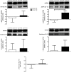
Systemic lupus erythematosus (SLE) patients may show increased insulin resistance (IR) when compared with their healthy peers. Exercise training has been shown to improve insulin sensitivity in other insulin-resistant populations, but it has never been tested in SLE. Therefore, the aim of the present study was to assess the efficacy of a moderate-intensity exercise training program on insulin sensitivity and potential underlying mechanisms in SLE patients with mild/inactive disease. A 12-week, randomized controlled trial was conducted. Nineteen SLE patients were randomly assigned into two groups: trained (SLE-TR, n = 9) and non-trained (SLE-NT, n = 10). Before and after 12 weeks of the exercise training program, patients underwent a meal test (MT), from which surrogates of insulin sensitivity and beta-cell function were determined. Muscle biopsies were performed after the MT for the assessment of total and membrane GLUT4 and proteins related to insulin signaling [Akt and AMP-activated protein kinase (AMPK)]. SLE-TR showed, when compared with SLE-NT, significant decreases in fasting insulin [-39 vs. +14%, p = 0.009, effect size (ES) = -1.0] and in the insulin response to MT (-23 vs. +21%, p = 0.007, ES = -1.1), homeostasis model assessment IR (-30 vs. +15%, p = 0.005, ES = -1.1), a tendency toward decreased proinsulin response to MT (-19 vs. +6%, p = 0.07, ES = -0.9) and increased glucagon response to MT (+3 vs. -3%, p = 0.09, ES = 0.6), and significant increases in the Matsuda index (+66 vs. -31%, p = 0.004, ES = 0.9) and fasting glucagon (+4 vs. -8%, p = 0.03, ES = 0.7). No significant differences between SLT-TR and SLT-NT were observed in fasting glucose, glucose response to MT, and insulinogenic index (all p > 0.05). SLE-TR showed a significant increase in AMPK Thr 172 phosphorylation when compared to SLE-NT (+73 vs. -12%, p = 0.014, ES = 1.3), whereas no significant differences between groups were observed in Akt Ser 473 phosphorylation, total and membrane GLUT4 expression, and GLUT4 translocation (all p > 0.05). In conclusion, a 12-week moderate-intensity aerobic exercise training program improved insulin sensitivity in SLE patients with mild/inactive disease. This effect appears to be partially mediated by the increased insulin-stimulated skeletal muscle AMPK phosphorylation.
Clinical trial registration: www.ClinicalTrials.gov, identifier NCT01515163.
Keywords: GLUT4; aerobic exercise; glucagon; inflammatory rheumatic disease; insulin resistance.
Figures


References
-
- Yang G, Li C, Gong Y, Fang F, Tian H, Li J, et al. Assessment of insulin resistance in subjects with normal glucose tolerance, hyperinsulinemia with normal blood glucose tolerance, impaired glucose tolerance, and newly diagnosed type 2 diabetes (prediabetes insulin resistance research). J Diabetes Res (2016) 2016:9270768.10.1155/2016/9270768 - DOI - PMC - PubMed
Publication types
MeSH terms
Substances
Associated data
LinkOut - more resources
Full Text Sources
Other Literature Sources
Medical
Molecular Biology Databases
Research Materials

