

Susceptibility MRI captures nigral pathology in patients with parkinsonian syndromes
- PMID: 29756231
- PMCID: PMC6185787
- DOI: 10.1002/mds.27381
Susceptibility MRI captures nigral pathology in patients with parkinsonian syndromes
Abstract
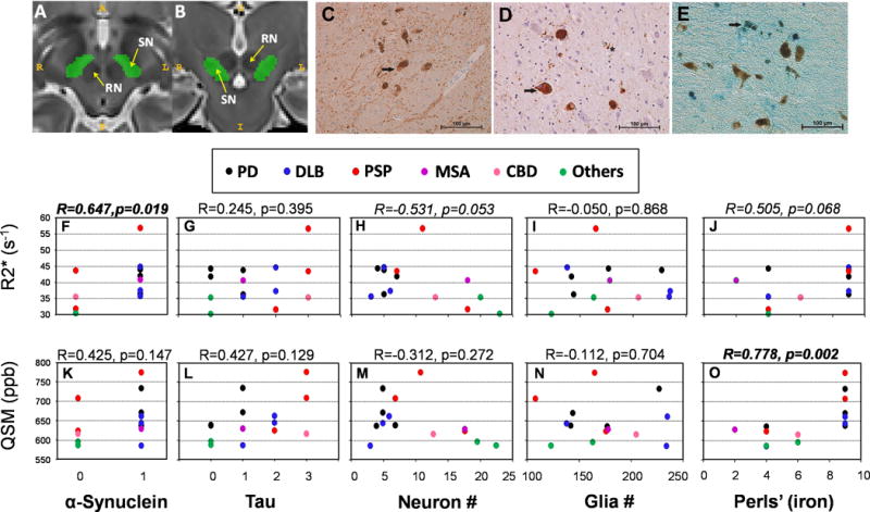
Background: Parkinsonisms are neurodegenerative disorders characterized pathologically by α-synuclein-positive (e.g., PD, diffuse Lewy body disease, and MSA) and/or tau-positive (e.g., PSP, cortical basal degeneration) pathology. Using R2* and quantitative susceptibility mapping, susceptibility changes have been reported in the midbrain of living parkinsonian patients, although the exact underlying pathology of these alterations is unknown.
Objective: The current study investigated the pathological correlates of these susceptibility MRI measures.
Methods: In vivo MRIs (T1- and T2-weighted, and T2*) and pathology were obtained from 14 subjects enrolled in an NINDS PD Biomarker Program (PDBP). We assessed R2* and quantitative susceptibility mapping values in the SN, semiquantitative α-synuclein, tau, and iron values, as well as neuronal and glial counts. Data were analyzed using age-adjusted Spearman correlations.
Results: R2* was associated significantly with nigral α-synuclein (r = 0.746; P = 0.003). Quantitative susceptibility mapping correlated significantly with Perls' (r = 0.758; P = 0.003), but not with other pathological measurements. Neither measurement correlated with tau or glial cell counts (r ≤ 0.11; P ≥ 0.129).
Conclusions: Susceptibility MRI measurements capture nigral pathologies associated with parkinsonian syndromes. Whereas quantitative susceptibility mapping is more sensitive to iron, R2* may reflect pathological aspects of the disorders beyond iron such as α-synuclein. They may be invaluable tools in diagnosing differential parkinsonian syndromes, and tracking in living patients the dynamic changes associated with the pathological progression of these disorders. © 2018 International Parkinson and Movement Disorder Society.
Keywords: iron; substantia nigra; susceptibility MRI; tau; α-synuclein.
© 2018 International Parkinson and Movement Disorder Society.
Conflict of interest statement
Figures
References
-
- Li K, Reichmann H. Role of iron in neurodegenerative diseases. J Neural Transm (Vienna) 2016;123:389–399. - PubMed
-
- Riederer P, Sofic E, Rausch WD, et al. Transition metals, ferritin, glutathione, and ascorbic acid in parkinsonian brains. J Neurochem. 1989;52:515–520. - PubMed
-
- Sofic E, Riederer P, Heinsen H, et al. Increased iron (III) and total iron content in post mortem substantia nigra of parkinsonian brain. J Neural Transm. 1988;74:199–205. - PubMed
Publication types
MeSH terms
Substances
Grants and funding
LinkOut - more resources
Full Text Sources
Other Literature Sources
Medical
Miscellaneous
