. 2018 Jun 20;46(11):5717-5725.
doi: 10.1093/nar/gky387.

Modulation of H-NS transcriptional silencing by magnesium

Affiliations

Modulation of H-NS transcriptional silencing by magnesium

W Ryan Will et al. Nucleic Acids Res. .

Abstract

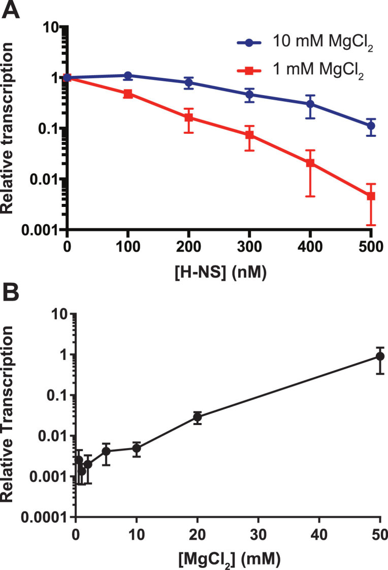
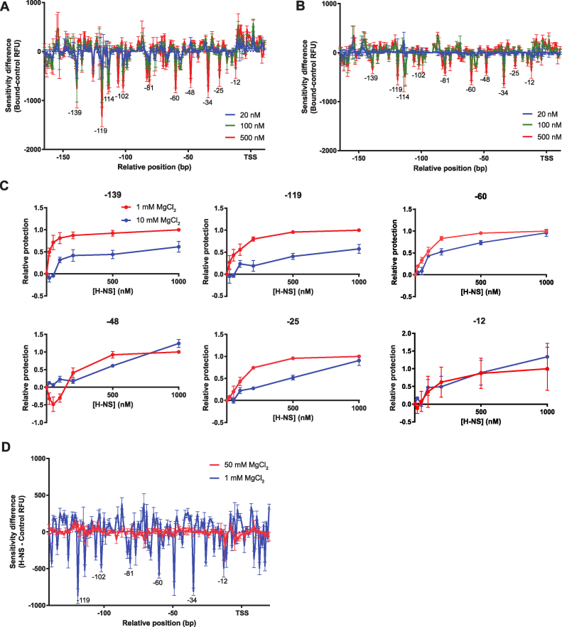
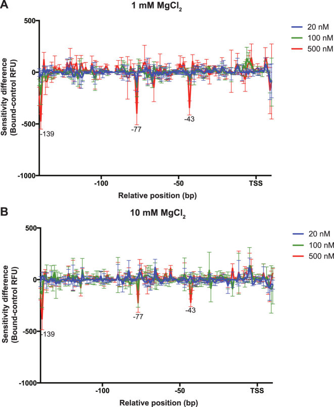
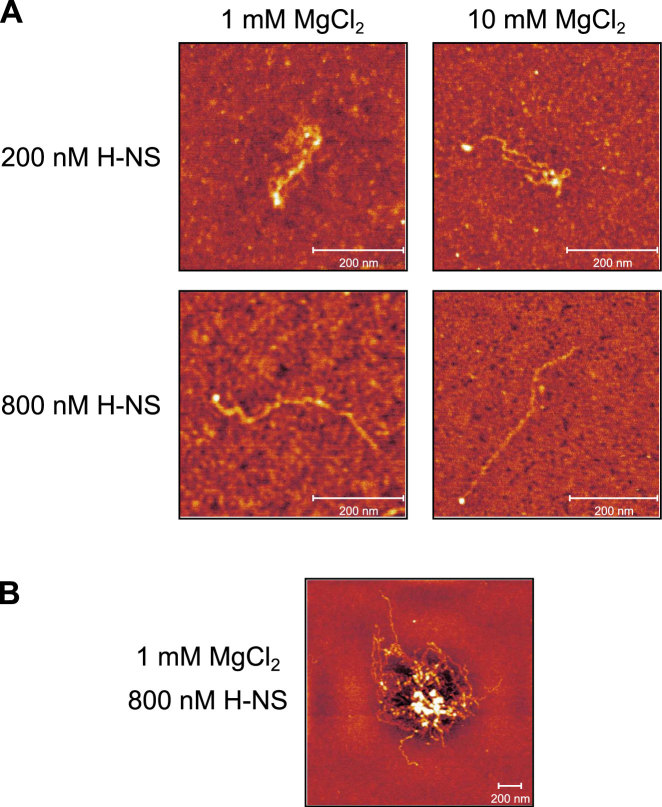
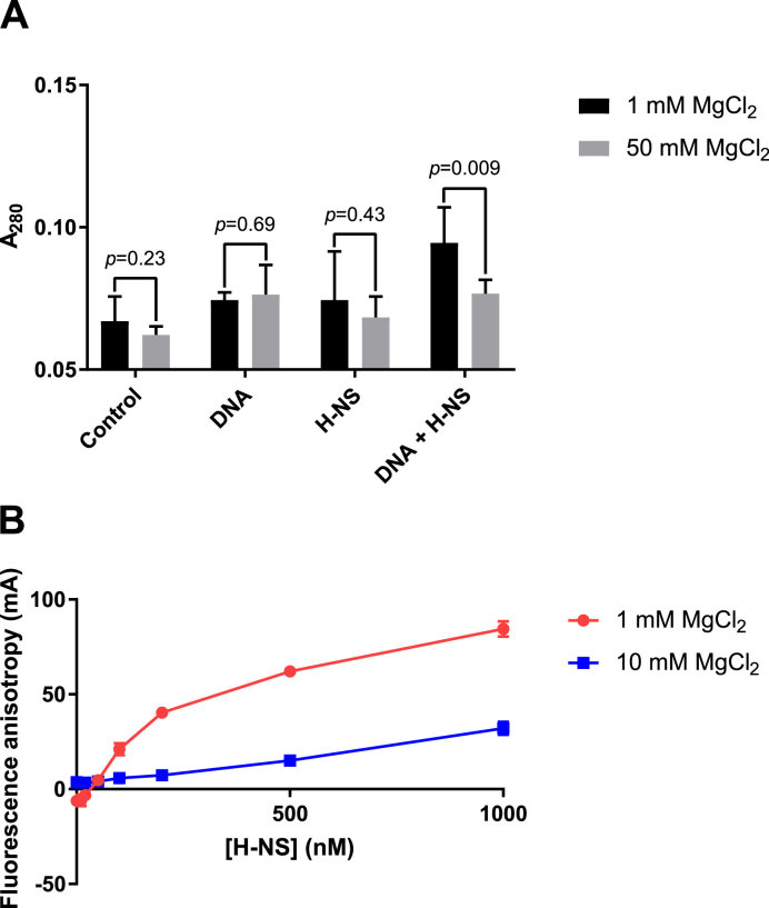
The bacterial histone-like protein H-NS silences AT-rich DNA, binding DNA as 'stiffened' filaments or 'bridged' intrastrand loops. The switch between these modes has been suggested to depend on the concentration of divalent cations, in particular Mg2+, with elevated Mg2+ concentrations associated with a transition to bridging. Here we demonstrate that the observed binding mode is a function of the local concentration of H-NS and its cognate binding sites, as well as the affinity of the interactions between them. Mg2+ does not control a binary switch between these two modes but rather modulates the affinity of this interaction, inhibiting the DNA-binding and silencing activity of H-NS in a continuous linear fashion. The direct relationship between conditions that favor stiffening and transcriptional silencing activity suggests that although both modes can occur in the cell, stiffening is the predominant mode of binding at silenced genes.


Figures

Figure 1.
Figure 1.
H-NS-mediated silencing is inhibited by increasing Mg2+ concentrations. (A) IVT was performed on pagC template with increasing concentrations of H-NS under either stiffening (1 mM MgCl2; red line) or bridging (10 mM MgCl2; blue line) conditions. Reactions are normalized to the 0 nM reaction. To determine if H-NS exhibits a binary response to Mg2+, IVT was performed on pagC template in the presence of 500nM H-NS and increasing concentrations of MgCl2 (B). Transcript quantities were normalized to an H-NS-free control reaction for each concentration of MgCl2 (see Supplementary Figure S3 for pre-normalization and control data). Data represent the mean ± SEM; n = 3.
Figure 2.
Figure 2.
H-NS binding is inhibited by increasing Mg2+ concentrations. DNase I Differential DNA Footprint Analysis was performed on the pagC promoter region with increasing concentrations of H-NS under either stiffening (A; 1 mM MgCl2) or bridging (B; 10 mM MgCl2) conditions. Results are presented as DDFA plots, representing the difference in fluorescent peak height (relative fluorescence units; RFU) between the control and H-NS-bound samples at three different H-NS concentrations (20 nM; blue line, 100 nM; green line, and 500 nM; red line). The approximate position of nucleotides relative to the TSS is indicated on the horizontal axis. Data represent the mean ± SD; n = 3. Downward valleys are regions of protection from DNase I digestion, indicating protein binding, whereas upward peaks indicate regions of increased DNase I sensitivity, suggesting bending or distortion of the DNA duplex. See Supplementary Figure S4 for representative electropherograms. (C) The fractional protection of several bases (–139, –119, –60, –48, –25 and –12 relative to the TSS) was determined relative to the reaction conditions resulting in maximum protection (1000 nM H-NS and 1 mM MgCl2) and plotted for a range of H-NS concentrations in either 1 mM (red line) or 10 mM (blue line) MgCl2, indicating the approximate affinity of the interaction between each base and H-NS. (D) 50 mM MgCl2 completely inhibited H-NS binding. DNase I DDFA was performed on the pagC promoter region in the presence of 500 nM H-NS and either 1 mM (blue line) or 50 mM (red line) MgCl2.
Figure 3.
Figure 3.
Local DNA structure is similar at low and high Mg2+ concentrations. UV laser Differential DNA Footprinting Analysis was performed on the pagC promoter region in increasing concentrations of H-NS under either stiffening (A; 1 mM MgCl2) or bridging (B; 10 mM MgCl2) conditions. Results are presented as DDFA plots, representing the difference in fluorescent peak height (relative fluorescence units; RFU) between control and H-NS-bound samples at three different H-NS concentrations (20 nM; blue line, 100 nM; green line and 500 nM; red line). The approximate position of cross-linked nucleotides relative to the TSS is indicated on the horizontal axis. Data represent the mean ± SD; n = 3. Significant cross-linking, suggestive of structural changes, is detectable at positions –139, –77 and –43. Representative electropherograms are shown in Supplementary Figure S7.
Figure 4.
Figure 4.
H-NS binding, but not binding mode, correlates with Mg2+ concentrations. The linearized 2.7 kb (∼610 nm) pagC region was incubated in the presence of 1 or 10 mM MgCl2, and 200 or 800 nM H-NS, at room temperature, and applied to glutaraldehyde-treated mica. H-NS-pagC complexes were then imaged by AFM in air. Representative discrete complexes are shown (A). At a concentration of 200 nM, which is sufficient for silencing (Figure 1), H-NS appears to form bridges in 1 mM MgCl2 (see Supplementary Figure S10 for additional images of the bridged complex, as well as the unbound DNA control), but stiffened filaments form at 800 nM H-NS. Similarly, in 10 mM MgCl2, H-NS binding is limited, but H-NS appears to form bridges when present at a concentration of 200 nM. However, filaments can be observed when the H-NS concentration is increased to 800 nM. Multimolecular aggregates, suggestive of bridging, can be observed under all binding conditions, but appear to be largest in 1 mM MgCl2 and 800 nM H-NS (B). Observed binding modes are scored in Supplementary Figure S11. Representative aggregates are shown for all conditions in Supplementary Figure S12A. The diameters of observed aggregates, which are reflective of H-NS binding and oligomerization, are indicated in Supplementary Figure S12B.
Figure 5.
Figure 5.
H-NS-mediated aggregation is dependent on DNA binding, which is inhibited by Mg2+. The linearized 2.7 kb pagC region was incubated in the presence of 800 nM H-NS and either 1 or 50 mM MgCl2. Light scattering, which is indicative of aggregate formation, was measured at 280 nm (A). An increase in light scattering was detected in samples containing both H-NS and DNA at 1 mM MgCl2. However, a significant increase in light scattering was not detected in 50 mM MgCl2, a concentration sufficient to inhibit DNA binding. This indicates that aggregation requires DNA binding, which is inhibited by MgCl2. Data represent the mean ± SD; n ≥ 5. P-values are indicated above each pair of samples. To confirm that MgCl2 inhibits DNA binding directly, fluorescence anisotropy was performed using a 36bp target containing a high affinity H-NS binding site (B). 6-FAM-labeled DNA was incubated in the presence of increasing concentrations of H-NS and 1 or 10 mM MgCl2, as indicated, and anisotropy, which is indicative of H-NS binding, was measured. Anisotropy is significantly decreased at 10 mM MgCl2, indicating that MgCl2 directly inhibits the H-NS–DNA interaction. Data represent the mean ± SD of three independent experiments, each consisting of three technical replicates. The anisotropy of the reaction buffer containing 1 mM MgCl2 is subtracted from all samples to correct for background fluorescence.

Similar articles

Cited by

References

    1. Navarre W.W., Porwollik S., Wang Y., McClelland M., Rosen H., Libby S.J., Fang F.C.. Selective silencing of foreign DNA with low GC content by the H-NS protein in Salmonella. Science. 2006; 313:236–238. - PubMed
    1. Lucchini S., Rowley G., Goldberg M.D., Hurd D., Harrison M., Hinton J.C.. H-NS mediates the silencing of laterally acquired genes in bacteria. PLoS Pathog. 2006; 2:e81. - PMC - PubMed
    1. Will W.R., Navarre W.W., Fang F.C.. Integrated circuits: how transcriptional silencing and counter-silencing facilitate bacterial evolution. Curr. Opin. Microbiol. 2015; 23:8–13. - PMC - PubMed
    1. Gordon B.R., Li Y., Cote A., Weirauch M.T., Ding P., Hughes T.R., Navarre W.W., Xia B., Liu J.. Structural basis for recognition of AT-rich DNA by unrelated xenogeneic silencing proteins. Proc. Natl. Acad. Sci. U.S.A. 2011; 108:10690–10695. - PMC - PubMed
    1. Stella S., Spurio R., Falconi M., Pon C.L., Gualerzi C.O.. Nature and mechanism of the in vivo oligomerization of nucleoid protein H-NS. EMBO J. 2005; 24:2896–2905. - PMC - PubMed

Publication types