

Interactions between lysyl oxidases and ADAMTS proteins suggest a novel crosstalk between two extracellular matrix families
- PMID: 29758265
- PMCID: PMC6240406
- DOI: 10.1016/j.matbio.2018.05.003
Interactions between lysyl oxidases and ADAMTS proteins suggest a novel crosstalk between two extracellular matrix families
Abstract
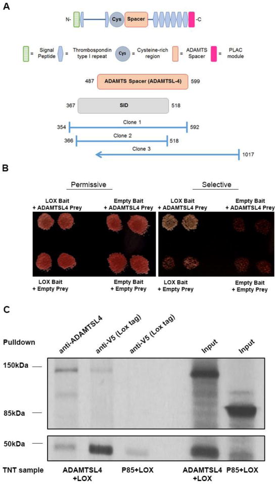
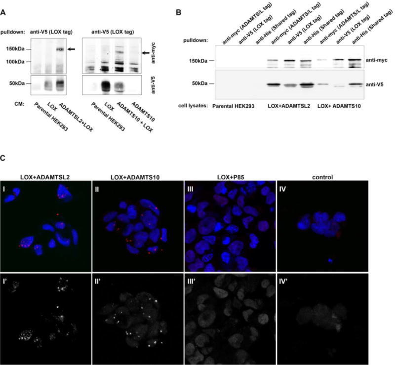
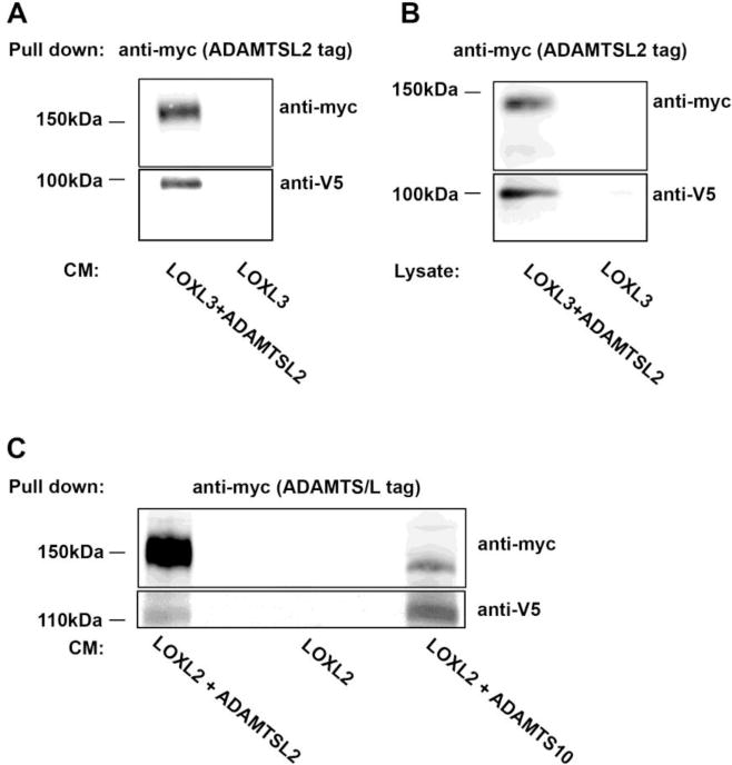
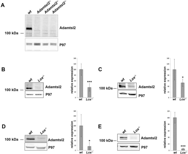
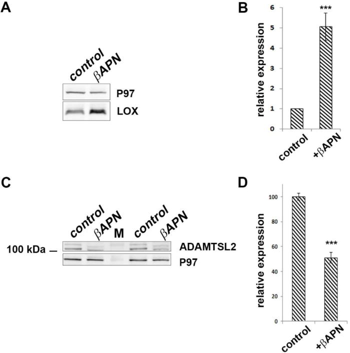
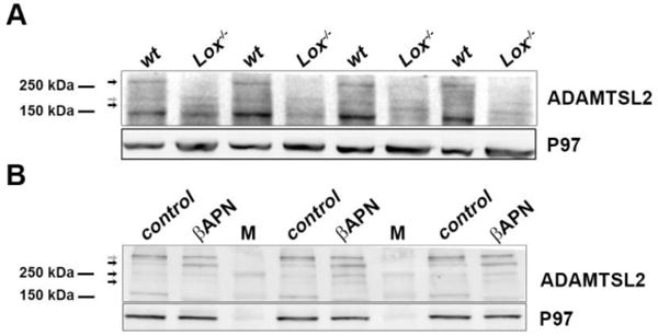
The extracellular matrix (ECM) regulates numerous cellular events in addition to providing structural integrity. Among several protein and enzyme families implicated in functions of the ECM, the lysyl oxidases and ADAMTS proteins are known to participate in microfibril and elastic fiber formation as well as ECM-associated signaling. A yeast two-hybrid screen to identify lysyl oxidase (LOX) binding proteins identified ADAMTSL4 as a potential interactor. We demonstrate here that several members of the LOX and ADAMTS families interact with one another. Upon investigating the interaction between LOX and ADAMTSL2 we found that the absence or inhibition of Lox affected ADAMTSL2 molecular forms and reduced its tissue levels. Thus, ADAMTSL2 stability and inter-molecular complexes may depend on the activity of lysyl oxidases.
Copyright © 2018 International Society of Matrix Biology. Published by Elsevier B.V. All rights reserved.
Conflict of interest statement
The authors declare no competing or financial interests.
Figures
Similar articles
-
Vascular lysyl oxidase over-expression alters extracellular matrix structure and induces oxidative stress.Clin Investig Arterioscler. 2017 Jul-Aug;29(4):157-165. doi: 10.1016/j.arteri.2017.01.004. Epub 2017 Jun 16. Clin Investig Arterioscler. 2017. PMID: 28624291 English, Spanish.
-
Lysyl oxidases: from enzyme activity to extracellular matrix cross-links.Essays Biochem. 2019 Sep 13;63(3):349-364. doi: 10.1042/EBC20180050. Print 2019 Sep 13. Essays Biochem. 2019. PMID: 31488698 Review.
-
Differential expression of lysyl oxidases LOXL1 and LOX during growth and aging suggests specific roles in elastin and collagen fiber remodeling in rat aorta.Rejuvenation Res. 2008 Oct;11(5):883-9. doi: 10.1089/rej.2008.0760. Rejuvenation Res. 2008. PMID: 18803461
-
New insights into elastic fiber assembly.Birth Defects Res C Embryo Today. 2007 Dec;81(4):229-40. doi: 10.1002/bdrc.20111. Birth Defects Res C Embryo Today. 2007. PMID: 18228265 Review.
-
Perturbations to lysyl oxidase expression broadly influence the transcriptome of lung fibroblasts.Physiol Genomics. 2017 Aug 1;49(8):416-429. doi: 10.1152/physiolgenomics.00026.2017. Epub 2017 Jul 10. Physiol Genomics. 2017. PMID: 28698228
Cited by
-
Ovariectomy drives increase of an ECM transcription signature in the posterior eye and retina.Vision Res. 2024 Dec;225:108507. doi: 10.1016/j.visres.2024.108507. Epub 2024 Oct 30. Vision Res. 2024. PMID: 39476526
-
ADAMTSL2 gene variant in patients with features of autosomal dominant connective tissue disorders.Am J Med Genet A. 2021 Mar;185(3):743-752. doi: 10.1002/ajmg.a.62030. Epub 2020 Dec 27. Am J Med Genet A. 2021. PMID: 33369194 Free PMC article.
-
Spatial and Single-Cell Transcriptomics Reveals the Regional Division of the Spatial Structure of MASH Fibrosis.Liver Int. 2025 Apr;45(4):e16125. doi: 10.1111/liv.16125. Epub 2024 Oct 14. Liver Int. 2025. PMID: 39400982 Free PMC article.
-
Biomarker discovery for chronic liver diseases by multi-omics - a preclinical case study.Sci Rep. 2020 Jan 28;10(1):1314. doi: 10.1038/s41598-020-58030-6. Sci Rep. 2020. PMID: 31992752 Free PMC article.
-
The potential prognostic values of the ADAMTS-like protein family: an integrative pan-cancer analysis.Ann Transl Med. 2021 Oct;9(20):1562. doi: 10.21037/atm-21-4946. Ann Transl Med. 2021. PMID: 34790768 Free PMC article.
References
-
- Csiszar K. Lysyl oxidases: a novel multifunctional amine oxidase family. Prog Nucleic Acid Res Mol Biol. 2001;70:1–32. - PubMed
-
- Mäki JM. Lysyl oxidases in mammalian development and certain pathological conditions. Histol Histopathol. 2009;24(5):651–60. - PubMed
-
- Dawson DA, Rinaldi AC, Poch G. Biochemical and toxicological evaluation of agent-cofactor reactivity as a mechanism of action for osteolathyrism. Toxicology. 2002;177(2–3):267–84. - PubMed
-
- Guo DC, Regalado ES, Gong L, Duan X, Santos-Cortez RL, Arnaud P, Ren Z, Cai B, Hostetler EM, Moran R, Liang D, Estrera A, Safi HJ, Leal SM, Bamshad MJ, Shendure J, Nickerson DA, Jondeau G, Boileau C, Milewicz DM. LOX Mutations Predispose to Thoracic Aortic Aneurysms and Dissections. Circ Res. 2016;118(6):928–34. - PMC - PubMed
Publication types
MeSH terms
Substances
Grants and funding
LinkOut - more resources
Full Text Sources
Other Literature Sources
Molecular Biology Databases
Research Materials
Miscellaneous
