Review
. 2018 Nov:67:150-167.
doi: 10.1016/j.preteyeres.2018.05.002. Epub 2018 Jul 13.

Pathogenesis of Keratoconus: The intriguing therapeutic potential of Prolactin-inducible protein

Affiliations
Review

Pathogenesis of Keratoconus: The intriguing therapeutic potential of Prolactin-inducible protein

Rabab Sharif et al. Prog Retin Eye Res. 2018 Nov.

Abstract

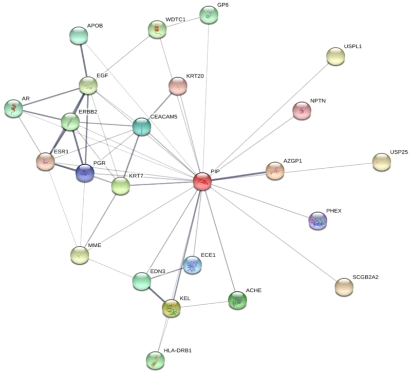
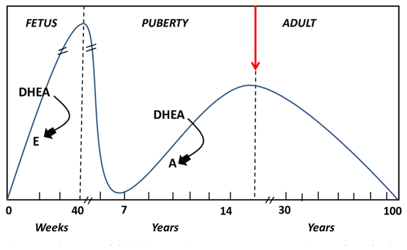
Keratoconus (KC) is the most common ectatic corneal disease, with clinical findings that include discomfort, visual disturbance and possible blindness if left untreated. KC affects approximately 1:400 to 1:2000 people worldwide, including both males and females. The aetiology and onset of KC remains a puzzle and as a result, the ability to treat or reverse the disease is hampered. Sex hormones are known to play a role in the maintenance of the structure and integrity of the human cornea. Hormone levels have been reported to alter corneal thickness, curvature, and sensitivity during different times of menstrual cycle. Surprisingly, the role of sex hormones in corneal diseases and KC has been largely neglected. Prolactin-induced protein, known to be regulated by sex hormones, is a new KC biomarker that has been recently proposed. Studies herein discuss the role of sex hormones as a control mechanism for KC onset and progression and evidence supporting the view that prolactin-induced protein is an important hormonally regulated biomarker in KC is discussed.

Keywords: Bodily fluids; Human cornea; Keratoconus; Prolactin-induced protein; Sex hormones.


Conflict of interest statement

Conflicts of interest

Authors declare no conflict of interest.

Figures

Fig. 1.
Fig. 1.
The layers of the human cornea. Illustrative image of the five human corneal layers, from top to bottom: corneal epithelium, Bowman’s layer, corneal stroma, Descemet’s membrane, and corneal endothelium. Image proportions not to scale.
Fig. 2.
Fig. 2.
Slit lamp images of KC signs. A: Corneal thinning at the apex of the cone. B: Corneal ectasia, with indentation of the inferior eyelid upon down gazing C: Paracentral stromal scars. D: Fleischer’s ring, a pigmented, often incomplete line of iron deposits running around the base of the cone. E: Vogt’s stria: fine vertical lines, which are breaks in the deep stroma and Descemet’s membrane. F: Corneal hydrops, the most acute presentation of KC. Diffuse stromal opacity and edema, caused by breaks in the Descemet’s membrane leading to influx of fluid in the stroma.
Fig. 3.
Fig. 3.
Anterior cross-section Optical Coherence Tomography (OCT). A1 and B1 show the scan location on the cornea. A2 and B2 show the cross-section view of the cornea. A: Healthy cornea. B: Severe KC cornea, characteristic protrusion and thinning as well as scarring at the top of the cone.
Fig. 4.
Fig. 4.
Four image tomography map from Pentacam® HR. A: Healthy cornea B: KC with severe ectasia and thinning, sagittal curvature (upper right), anterior elevation subtraction maps (upper left), corneal thickness (lower right) and posterior elevation subtraction map (lower left) are shown.
Fig. 5.
Fig. 5.
Contact lenses and Intra-corneal ring segments (ICRS). A: Rigid-gas permeable lens B: Hybrid lens with rigid center and soft periphery. Notice transition between rigid and soft (arrow) C: Scleral contact lens. D: Lenses A-C out of eye. E: ICRS. Notice deposit on inner arc (arrow).
Fig. 6.
Fig. 6.
Illustrative image of our 3-D in vitro model. Cells are harvested from donor corneas and grown on polycarbonate membranes with vitamin C stimulation.
Fig. 7.
Fig. 7.
Quantitative Polymerase Chain Reaction (QT-PCR) showing significant upregulation in ER (ESR1; Fig. 7A; p < 0.05) and a downregulation of AR (AR; Fig. 7B; p < 0.05) in HKCs when compared to HCFs. Data shown is preliminary.
Fig. 8.
Fig. 8.
Quantification of Collagen type III (Col III) and cellular Fibronectin (cFN) protein levels in HCF and HKC 3-D constructs. (A) Col III was significantly upregulated in HKCs following exogenous DHEA (p < 0.05). Col III was significantly downregulated in HCFs following exogenous estrone (p < 0.01). (B) cFN was significantly downregulated in HKCs following exogenous DHEA (p < 0.05). cFN was significantly downregulated in both HCFs and HKCs following exogenous Estrone (p < 0.01). Values normalized to HCFs (n = 4). Data shown is preliminary.
Fig. 9.
Fig. 9.
Mechanistic network derived from differential gene expression profile for three male and three female donors for HCFs (n = 6) and HKCs (n = 6), using whole-transcriptome high-throughput sequencing (RNA-Seq). Network shows genes (nodes) connected by lines (edges) indicating a known relationship/interaction in the IPA Knowledge Base. Data shown is preliminary.
Fig. 10.
Fig. 10.
Structure of the Prolactin-Induced Protein based PyMOL software (Hassan et al., 2008).
Fig. 11.
Fig. 11.
Network view of the predicted interactors of Prolactin-Induced Protein: STRING database, illustrates the display of Protein-Protein interactions through a Java applet.
Fig. 12.
Fig. 12.
Quantification of PIP protein levels. (A) Human tear samples from healthy and KC donors (n = 32). (B) Human saliva samples from healthy and KC donors (n = 64). (C) HCFs and HKCs in 3-D constructs (n = 6). PIP was significantly downregulated in KC samples, compared to healthy controls, in all three systems; Tears (p < 0.0001), Saliva (p < 0.001), and Cells (p < 0.005).
Fig. 13.
Fig. 13.
Schematic of dehydroepiandrosterone (DHEA) production from fetal to adult life. The arrows indicate temporal changes in sex hormones controlling DHEA (E: estrogen, A: androgen).

References

    1. Abu-Amero, Helwa, Al-Muammar, Strickland, Hauser, Allingham, Liu, 2015. Screening of the seed region of Mir184 in keratoconus patients from Saudi Arabia. BioMed. Res. Int 2015, 604508. - PMC - PubMed
    1. Amsler M, 1946. Kératocône classique et kératocône fruste; arguments unitaires. Ophthalmologica 111, 96–101. - PubMed
    1. Annibalini G, Agostini D, Calcabrini C, Martinelli C, Colombo E, Guescini M, Tibollo P, Stocchi V, Sestili P, 2014. Effects of sex hormones on inflammatory response in male and female vascular endothelial cells. J. Endocrinol. Invest 37, 861–869. - PubMed
    1. Anwar M, Teichmann KD, 2002. Deep lamellar keratoplasty: surgical techniques for anterior lamellar keratoplasty with and without baring of Descemet’s membrane. Cornea 21, 374–383. - PubMed
    1. Atilano SR, Coskun P, Chwa M, Jordan N, Reddy V, Le K, Wallace DC, Kenney MC, 2005. Accumulation of mitochondrial DNA damage in keratoconus corneas. Invest. Ophthalmol. Vis. Sci 46, 1256–1263. - PubMed

Publication types