

Comprehensive characterization of circulating and bone marrow-derived multiple myeloma cells at minimal residual disease
- PMID: 29759150
- PMCID: PMC6145140
- DOI: 10.1053/j.seminhematol.2018.02.010
Comprehensive characterization of circulating and bone marrow-derived multiple myeloma cells at minimal residual disease
Abstract
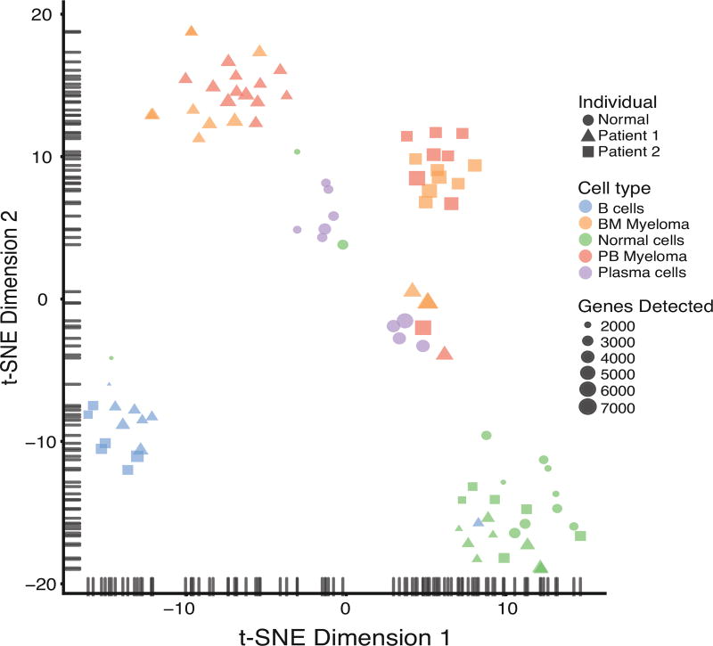
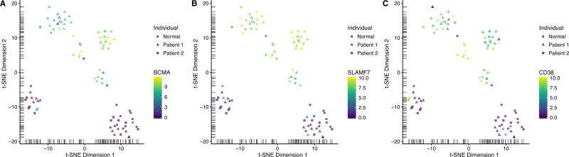
The presence or absence of minimal residual disease (MRD) in patients with multiple myeloma (MM) has emerged as a useful marker to determine the depth of remission. MRD negativity as an endpoint has been shown to be associated with improved progression-free survival in many studies. MRD detection is therefore part of numerous clinical trial protocols for MM. At the present time, two methodologies are most widely accepted for MRD detection: (1) multicolor flow cytometry and (2) next-generation sequencing-based clonotype detection. While both of those methodologies enable accurate quantification of MRD in the bone marrow (BM), with sensitivity as low as 10-5 to 10-6, there are several limitations to these methods. First, these approaches reveal the presence or absence of MRD but provide limited molecular information about MM. More comprehensive characterization of MM cells at the MRD stage may identify molecular mechanisms of drug resistance. Second, MRD detection in the BM is typically performed at one time point only, but more frequent detection may define the duration of the MRD status and thus refine its prognostic value. Third, less-invasive approaches that avoid the discomfort and risk associated with BM biopsy would be highly desirable, especially in elderly or frail patients. "Liquid biopsy" for the detection and characterization of circulating MM cells may address these issues. Although MRD detection in the peripheral blood at the same sensitivity as in the BM may be challenging, the identification of patients who do not achieve MRD negativity might reduce the need for BM biopsies. Here, we give an overview of approaches that have been described to detect and characterize MM cells when they occur at very low frequencies in the peripheral blood or in the BM, emphasizing recently described next-generation sequencing approaches for more comprehensive characterization of circulating MM cells.
Keywords: Circulating multiple myeloma cells; Clonal evolution; MRD; Next-generation sequencing.
Copyright © 2018 Elsevier Inc. All rights reserved.
Conflict of interest statement
All authors declare no relevant conflicts of interests.
Figures
References
-
- Hideshima T, Richardson P, Chauhan D, Palombella VJ, Elliott PJ, Adams J, et al. The proteasome inhibitor PS-341 inhibits growth, induces apoptosis, and overcomes drug resistance in human multiple myeloma cells. Cancer Res. 2001;61:3071–6. - PubMed
Publication types
MeSH terms
Grants and funding
LinkOut - more resources
Full Text Sources
Other Literature Sources
Medical
