. 2018 May 29;115(22):E4960-E4969.
doi: 10.1073/pnas.1801013115. Epub 2018 May 14.

Hydrogel delivery of lysostaphin eliminates orthopedic implant infection by Staphylococcus aureus and supports fracture healing

Affiliations

Hydrogel delivery of lysostaphin eliminates orthopedic implant infection by Staphylococcus aureus and supports fracture healing

Christopher T Johnson et al. Proc Natl Acad Sci U S A. .

Abstract

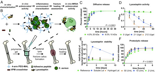
Orthopedic implant infections are a significant clinical problem, with current therapies limited to surgical debridement and systemic antibiotic regimens. Lysostaphin is a bacteriolytic enzyme with high antistaphylococcal activity. We engineered a lysostaphin-delivering injectable PEG hydrogel to treat Staphylococcus aureus infections in bone fractures. The injectable hydrogel formulation adheres to exposed tissue and fracture surfaces, ensuring efficient, local delivery of lysostaphin. Lysostaphin encapsulation within this synthetic hydrogel maintained enzyme stability and activity. Lysostaphin-delivering hydrogels exhibited enhanced antibiofilm activity compared with soluble lysostaphin. Lysostaphin-delivering hydrogels eradicated S. aureus infection and outperformed prophylactic antibiotic and soluble lysostaphin therapy in a murine model of femur fracture. Analysis of the local inflammatory response to infections treated with lysostaphin-delivering hydrogels revealed indistinguishable differences in cytokine secretion profiles compared with uninfected fractures, demonstrating clearance of bacteria and associated inflammation. Importantly, infected fractures treated with lysostaphin-delivering hydrogels fully healed by 5 wk with bone formation and mechanical properties equivalent to those of uninfected fractures, whereas fractures treated without the hydrogel carrier were equivalent to untreated infections. Finally, lysostaphin-delivering hydrogels eliminate methicillin-resistant S. aureus infections, supporting this therapy as an alternative to antibiotics. These results indicate that lysostaphin-delivering hydrogels effectively eliminate orthopedic S. aureus infections while simultaneously supporting fracture repair.

Keywords: S. aureus; biomaterials; infection; lysostaphin; orthopedics.


Conflict of interest statement

Conflict of interest statement: C.T.J. and A.J.G. are inventors on a patent application filed by the Georgia Tech Research Corp. based on the results in this study.

Figures

Fig. 1.
Fig. 1.
Lysostaphin-delivering hydrogel synthesis and characterization. (A) Outline of overall study design. (B) Schematic diagram of lysostaphin encapsulation within protease-degradable PEG-MAL hydrogel and subsequent application to infected femurs, which leads to fracture callus formation and healing. (C) Passive lysostaphin release with one-phase association fit with extra sum of squares F test to compare K values are different. (D) Optical density curves of lysostaphin-laden hydrogels placed in S. aureus UAMS-1 suspensions as a function of incubation time. (E) Lysostaphin activity as measured by the average half-life of the kinetic bacteria reduction assay (SI Appendix, Fig. S3 AD) at 1, 3, 7, and 14 d after hydrogel polymerization. (F) Protease-triggered release of lysostaphin with one-phase association fit using extra sum of squares F test to compare all K values are different. Lst, lysostaphin. Mean ± SD, n = 3–5.
Fig. 2.
Fig. 2.
Lysostaphin-laden hydrogels effectively kill bacteria in vitro. Bacterial counts reported as cfu per gel after 24 h of culture for (A) S. aureus Xen29, (B) S. aureus UAMS-1, (C) S. aureus 46106, and (D) S. epidermidis IDRL-8883. Mean ± SD, n = 3–4 per group. ****P < 0.0001, one-way ANOVA with Tukey’s post hoc test. (E and F) Biofilms were generated by culturing UAMS-1 for 24 h statically and were then treated overnight with a hydrogel or soluble enzyme. (E) Images and (F) quantification of average image intensity of live bacteria after treatment. One-way ANOVA with Holm–Sidak’s post hoc test between equivalent lysostaphin concentrations for hydrogel vs. soluble control. Mean ± SD, n = 3 per group. *P < 0.05, **P < 0.01. (Scale bar, 500 μm.)
Fig. 3.
Fig. 3.
Lysostaphin-delivering hydrogels eliminate bacteria in infected fractures. (A) Schematic diagram of mouse femur infection model. Quantification of S. aureus UAMS-1 recovered from the (B) tissue surrounding the femur, (C) femur bone, and (D) stabilization needle 7 d postfracture. Dashed line indicates detection limit. *P < 0.05, **P < 0.01. (E) Histological sections of femurs 7 d postfracture stained for H&E, Saf-O/FG, and Gram. Black arrows indicate gram-positive bacteria. Kruskal–Wallis test with Dunn’s multiple comparisons test. Ox., oxacillin; Sol., soluble. Mean ± SD n = 4–8, compilation of four independent experiments.
Fig. 4.
Fig. 4.
Lysostaphin-laden hydrogel therapy restores a sterile inflammatory environment. Femora were fractured and infected with UAMS-1 and treated with hydrogels with or without lysostaphin and the inflammatory milieu of tissue at the fracture site 7 d postinfection was assessed using multiplexed cytokine analysis. (A) Hierarchical cluster analysis of cytokine profiles using the Ward method. (B) Multivariate-ANOVA plot using a sum combination across cytokines, P < 0.001. (CK) Cytokines with statistically different tissue levels as determined using two-way ANOVA with a Bonferonni correction for multiple comparisons. U, UAMS-1; U + L, UAMS-1 + Lst; St, sterile. Mean ± SD, n = 6–8 per group. *P < 0.05, ***P < 0.001, ****P < 0.0001; ns, not significant.
Fig. 5.
Fig. 5.
Lysostaphin-delivering hydrogels enable fracture healing. (A) µCT reconstructions of the fracture callus 5 wk postoperation. (Scale bar, 1 mm.) Quantification of µCT reconstructions showing the (B) fracture callus volume and (C) bone volume within the fracture callus at 5 wk. (D) Mechanical strength of femurs as assessed by ex vivo torsion to failure testing. *P < 0.05, **P < 0.01, ***P < 0.001. (E) H&E, Saf-O/FG, and Gram staining of femurs. Black arrows indicate gram-positive bacteria. Kruskal–Wallis test with Dunn’s multiple comparisons test. Mean ± SD, n = 6–8, compilation of two independent experiments.
Fig. 6.
Fig. 6.
Lysostaphin-laden hydrogels clear MRSA infections. Quantification of MRSA USA300 recovered from the (A) tissue surrounding the femur, (B) femur, and (C) stabilization needle at 7 d postfracture. Dashed line indicates detection limit. ANOVA with Tukey’s post hoc test for A and B. Kruskal–Wallis test with Dunn’s multiple comparisons test for C. Mean ± SD, n = 3–4. *P < 0.05, ****P < 0.0001.

References

    1. Berríos-Torres SI, et al. Healthcare Infection Control Practices Advisory Committee Centers for disease control and prevention guideline for the prevention of surgical site infection, 2017. JAMA Surg. 2017;152:784–791. - PubMed
    1. Inzana JA, Schwarz EM, Kates SL, Awad HA. Biomaterials approaches to treating implant-associated osteomyelitis. Biomaterials. 2016;81:58–71. - PMC - PubMed
    1. Maradit Kremers H, et al. Prevalence of total hip and knee replacement in the United States. J Bone Joint Surg Am. 2015;97:1386–1397. - PMC - PubMed
    1. Antoci V, Chen AF, Parvizi J. 7.9 Orthopedic implant use and infection. In: Ducheyne P, editor. Comprehensive Biomaterials II. Elsevier; Oxford: 2017. pp. 133–151.
    1. Darouiche RO. Treatment of infections associated with surgical implants. N Engl J Med. 2004;350:1422–1429. - PubMed

Publication types

MeSH terms