. 2018 May 29;115(22):E5135-E5143.
doi: 10.1073/pnas.1802879115. Epub 2018 May 14.

Broad receptor engagement of an emerging global coronavirus may potentiate its diverse cross-species transmissibility

Affiliations

Broad receptor engagement of an emerging global coronavirus may potentiate its diverse cross-species transmissibility

Wentao Li et al. Proc Natl Acad Sci U S A. .

Abstract

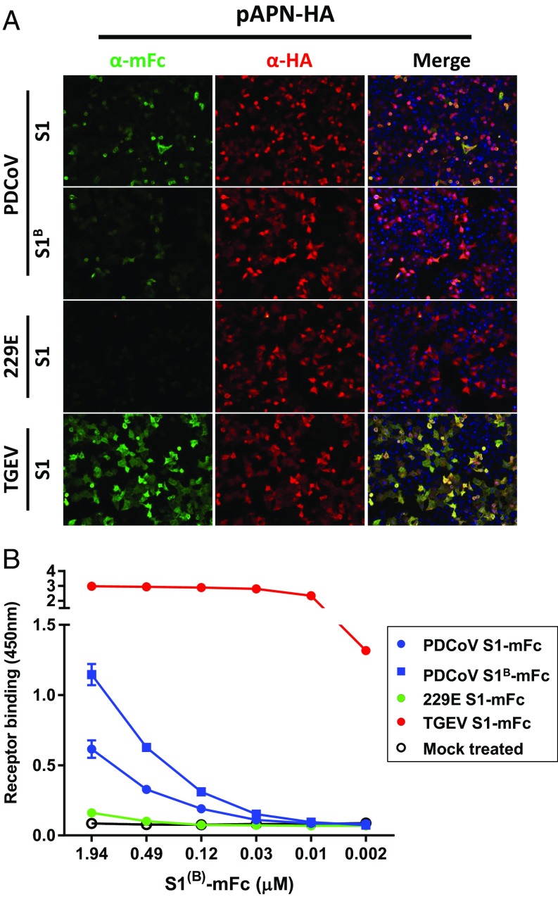
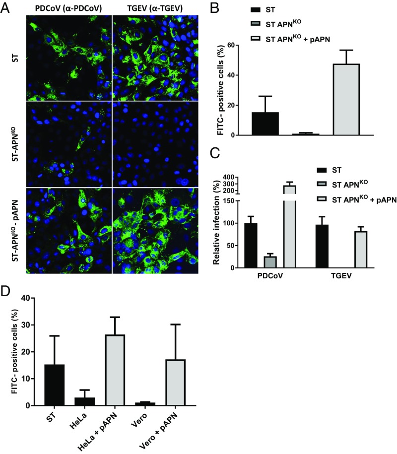
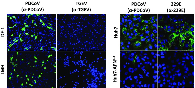
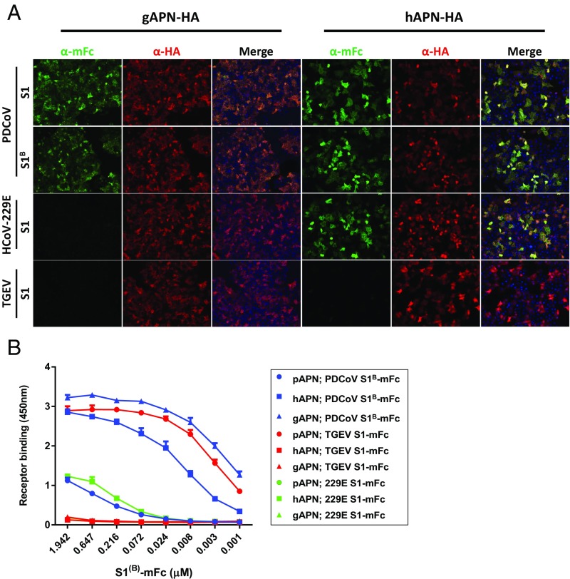
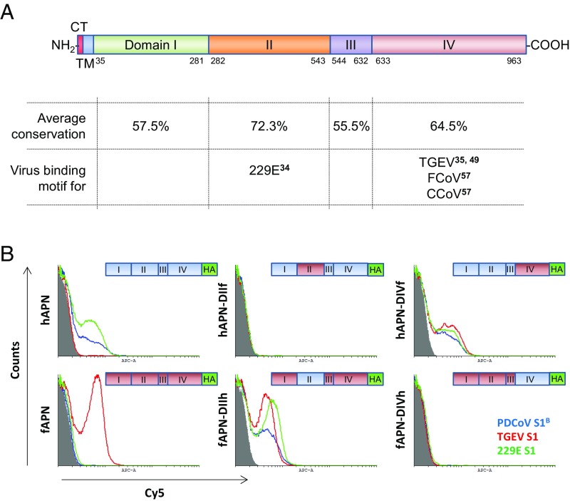
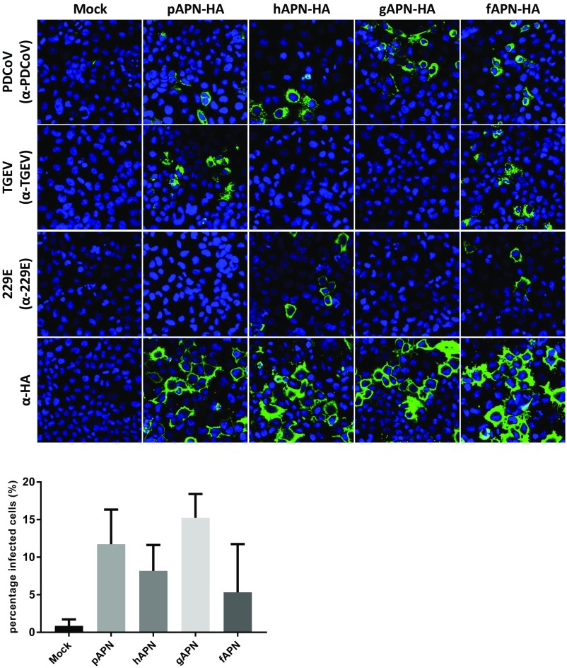
Porcine deltacoronavirus (PDCoV), identified in 2012, is a common enteropathogen of swine with worldwide distribution. The source and evolutionary history of this virus is, however, unknown. PDCoV belongs to the Deltacoronavirus genus that comprises predominantly avian CoV. Phylogenetic analysis suggests that PDCoV originated relatively recently from a host-switching event between birds and mammals. Insight into receptor engagement by PDCoV may shed light into such an exceptional phenomenon. Here we report that PDCoV employs host aminopeptidase N (APN) as an entry receptor and interacts with APN via domain B of its spike (S) protein. Infection of porcine cells with PDCoV was drastically reduced by APN knockout and rescued after reconstitution of APN expression. In addition, we observed that PDCoV efficiently infects cells of unusual broad species range, including human and chicken. Accordingly, PDCoV S was found to target the phylogenetically conserved catalytic domain of APN. Moreover, transient expression of porcine, feline, human, and chicken APN renders cells susceptible to PDCoV infection. Binding of PDCoV to an interspecies conserved site on APN may facilitate direct transmission of PDCoV to nonreservoir species, including humans, potentially reflecting the mechanism that enabled a virus, ancestral to PDCoV, to breach the species barrier between birds and mammals. The APN cell surface protein is also used by several members of the Alphacoronavirus genus. Hence, our data constitute the second identification of CoVs from different genera that use the same receptor, implying that CoV receptor selection is subjected to specific restrictions that are still poorly understood.

Keywords: APN; PDCoV; cross-species transmission; receptor; spike.


Conflict of interest statement

The authors declare no conflict of interest.

Figures

Fig. 1.
Fig. 1.
The S1 and the S1B domain of the PDCoV S protein bind to pAPN. (A) PDCoV S1 and S1B domain bind to HeLa cells overexpressing pAPN. HeLa cells mock-transfected or transfected with a plasmid encoding HA-tagged pAPN were incubated with equimolar amounts of PDCoV S1 or S1B proteins that were C-terminally tagged with the Fc domain of murine IgG2a (S1-mFc and S1B-mFc). Binding of the mFc-tagged S1 proteins and APN expression was measured by immunofluorescence assay using antibodies recognizing the mFc-tag or HA-tag, respectively. S1-mFc fusion proteins of the S proteins of TGEV (interacts with pAPN) and HCoV-229E (does not interact with pAPN) were taken along as controls. (Magnification: 200×.) (B) PDCoV S1 and S1B bind soluble pAPN. The 96-well plates coated with pAPN ectodomain were incubated with serially diluted PDCoV S1-mFc and S1B-mFc proteins. Bound S1(B)-mFc proteins were detected via HRP-conjugated antibodies recognizing the mFc-tag and subsequent development using HRP substrate. TGEV and HCoV-229E S1-mFc proteins were used as a positive and negative APN binding control, respectively. Error bars = SD; n = 2 (independent experiments each with two technical replicates).
Fig. 2.
Fig. 2.
Impact of APN expression on PDCoV infection. (A) PDCoV employs APN during ST cell entry. ST, ST-APNKO, and pAPN reconstituted knockout cells were inoculated with PDCoV or TGEV at a MOI of 1. Cells were fixed and permeabilized 16 hpi and stained using a PDCoV-specific antiserum or a TGEV-specific monoclonal antibody, respectively. Infection experiments were performed twice; representative pictures are shown. (Magnification: 300×.) (B) PDCoV infection levels on pAPN deficient and reconstituted ST cells were quantified by flow cytometry. Infection levels are shown normalized to parental ST cell infection. Experiments were repeated four times; averages are shown. (C) PDCoV and TGEV infection levels on pAPN-deficient and reconstituted ST cells quantified by counting virus-infected cells after immunofluorescent staining. Infection levels are shown relative to infection on the parental ST cells. Infection experiments were performed twice; a representative experiment is shown. (D) APN overexpression in HeLa and Vero cells potentiates PDCoV infection. HeLa and Vero cells stably expressing pAPN were generated by retroviral transduction. Cells were inoculated with PDCoV at a MOI = 1 (as titrated on ST cells) and infection levels were quantified at 16 hpi by flow cytometry. Experiments were performed two to five times. Average infection levels are shown relative to infection on the ST cells.
Fig. 3.
Fig. 3.
PDCoV infect cells of human and avian origin. Cell lines of avian (LMH and DF-1) and human (Huh7, Huh7-APNKO) origin were inoculated with PDCoV, TGEV, or HCoV-229E (MOI = 1) for 1 h, after which infection was assessed by immunostaining at 16 hpi. The results were confirmed in an independent experiment; pictures of a representative experiment are shown. (Magnification: Left, 200×; Right, 300×.)
Fig. 4.
Fig. 4.
PDCoV S1 binds to APN of human and avian origin. (A) PDCoV S1- and S1B bind to HeLa cells overexpressing galline and human APN. HeLa cells transiently transfected with a plasmid encoding HA-tagged human or gAPN (hAPN-HA and gAPN-HA) were incubated with equimolar amounts of Fc-tagged PDCoV S1 and S1B fusion proteins. Binding of the S1 proteins and APN expression was measured by immunofluorescence assay using antibodies recognizing the Fc-tag or HA-tag, respectively. Fc-tagged S1 proteins of the spike proteins of TGEV (binds pAPN) and HCoV-229E (does not bind pAPN) were used as controls. (Magnification: 200×.) (B) PDCoV S1 and S1B interact with human and galline APN. MaxiSorp-well plates coated with the ectodomain of porcine, human, or galline APN were incubated with serial dilutions of PDCoV S1-mFc and S1B-mFc proteins. Bound S1(B)-mFc proteins were detected via HRP-conjugated antibodies recognizing the mFc-tag and subsequent development using HRP substrate. TGEV and HCoV-229E S1-mFc proteins were used as a positive and negative APN binding control, respectively. Error bars = SD; n = 2 (independent experiments each with two technical replicates).
Fig. 5.
Fig. 5.
The highly conserved domain II of APN is engaged by PDCoV S domain S1B. (A) Schematic representation of the APN protein with the different domains and virus binding regions indicated. Schematic is based on the 963-amino acid sequence of pAPN (GenBank accession no. NP_999442.1) and was designed in accordance with the pAPN crystal structure as determined by Santiago et al. (75) (PDB ID code 5LDS). CT, cytosolic tail; TM, transmembrane domain. Virus binding regions of HCoV-229E, TGEV, feline coronavirus (FCoV), and canine coronavirus (CCoV) are indicated, as previously reported (34, 35, 49, 57). APN conservation across different classes of the subphylum Vertebrata was determined using APN amino acid sequences from mammals, amphibians, reptiles, birds, and fish. A complete alignment of the APN amino acid sequences used is shown in SI Appendix, Fig. S4. (B) PDCoV S1B interacts with the catalytic domain of APN (domain II). APN HeLa cells were transfected with plasmids encoding human, feline or chimeric APN proteins, and were examined the next day for interaction with PDCoV S1B, HCoV-229E S1, and TGEV S1-mFc fusion proteins by FACS analysis using a donkey α-mouse Cy5 conjugate (1:200). To control for cell surface expression, samples were stained with a rabbit α-HA antibody and corresponding conjugate (see SI Appendix, Fig. S7). S1(B)-mFc protein binding to cells is color-coded (PDCoV S1B, red; HCoV-229E, green; TGEV, blue) and compared with the HCoV-NL63 S1-mFc (negative control). Schematic representations of the C-terminally HA-tagged APN chimeras used in the assay are indicated in the plots. Data were analyzed using Cyflogic software.
Fig. 6.
Fig. 6.
PDCoV can use APN orthologs of nonhost species as a receptor. Orthologous APN molecules facilitate PDCoV entry into cells. (Upper) HeLa cells mock-transfected or transfected with plasmids encoding pAPN, hAPN, gAPN, and fAPN, were inoculated with PDCoV, HCoV-229E, or TGEV at a MOI = 1 for 1 h. Cells were fixed and permeabilized 24 hpi and stained with virus specific antibodies (PDCoV, TGEV, and HCoV-229E) or a mouse anti-HA antibody (Abcam, ab130275) and corresponding Alexa594 conjugate. Infection experiments were performed twice; pictures of a representative experiment are shown. (Magnification: 300×.) (Lower) Quantification of PDCoV infection. Amount of cells and PDCoV+ cells were counted using five or more pictures for each infection condition, and expressed relative to infection in mock-transfected HeLa cells.

Similar articles

Cited by

References

    1. Geoghegan JL, Duchêne S, Holmes EC. Comparative analysis estimates the relative frequencies of co-divergence and cross-species transmission within viral families. PLoS Pathog. 2017;13:e1006215. - PMC - PubMed
    1. Kitchen A, Shackelton LA, Holmes EC. Family level phylogenies reveal modes of macroevolution in RNA viruses. Proc Natl Acad Sci USA. 2011;108:238–243. - PMC - PubMed
    1. World Health Organization 2015 Summary of probable SARS cases with onset of illness from 1 November 2002 to 31 July 2003. Available at www.who.int/csr/sars/country/table2004_04_21/en/. Accessed January 29, 2018.
    1. Li W, et al. Receptor and viral determinants of SARS-coronavirus adaptation to human ACE2. EMBO J. 2005;24:1634–1643. - PMC - PubMed
    1. de Wit E, van Doremalen N, Falzarano D, Munster VJ. SARS and MERS: Recent insights into emerging coronaviruses. Nat Rev Microbiol. 2016;14:523–534. - PMC - PubMed

Publication types

MeSH terms

LinkOut - more resources