. 2018 Aug 23;132(8):837-848.
doi: 10.1182/blood-2017-11-814319. Epub 2018 May 14.

CTCF boundary remodels chromatin domain and drives aberrant HOX gene transcription in acute myeloid leukemia

Affiliations

CTCF boundary remodels chromatin domain and drives aberrant HOX gene transcription in acute myeloid leukemia

Huacheng Luo et al. Blood. .

Abstract

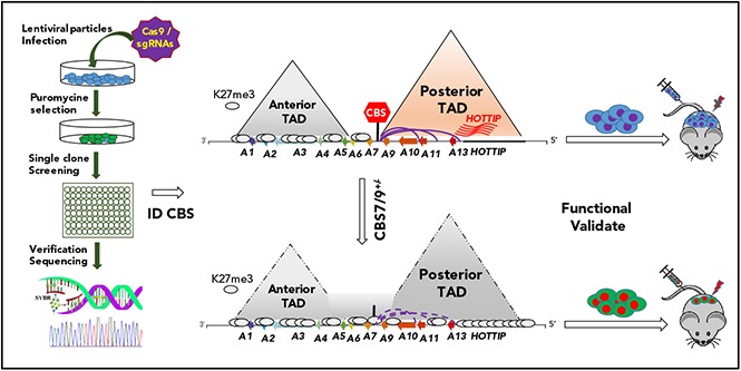
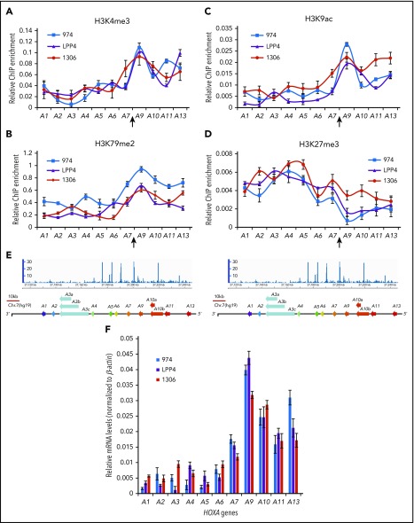
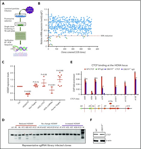
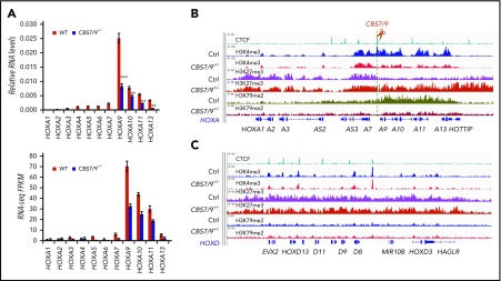
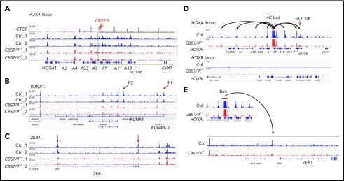
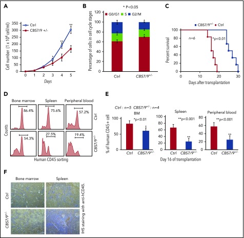
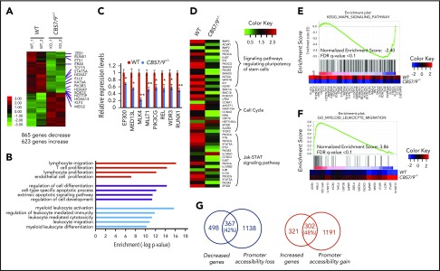
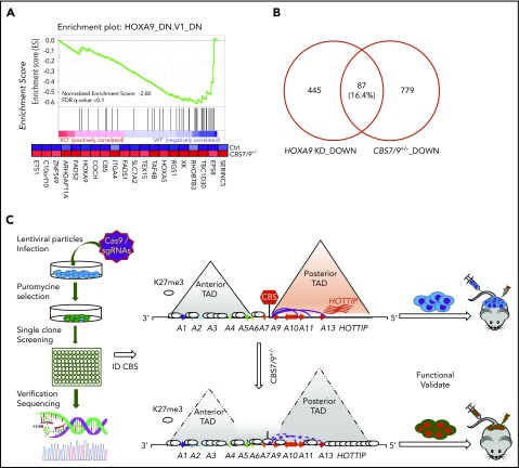
HOX gene dysregulation is a common feature of acute myeloid leukemia (AML). The molecular mechanisms underlying aberrant HOX gene expression and associated AML pathogenesis remain unclear. The nuclear protein CCCTC-binding factor (CTCF), when bound to insulator sequences, constrains temporal HOX gene-expression patterns within confined chromatin domains for normal development. Here, we used targeted pooled CRISPR-Cas9-knockout library screening to interrogate the function of CTCF boundaries in the HOX gene loci. We discovered that the CTCF binding site located between HOXA7 and HOXA9 genes (CBS7/9) is critical for establishing and maintaining aberrant HOXA9-HOXA13 gene expression in AML. Disruption of the CBS7/9 boundary resulted in spreading of repressive H3K27me3 into the posterior active HOXA chromatin domain that subsequently impaired enhancer/promoter chromatin accessibility and disrupted ectopic long-range interactions among the posterior HOXA genes. Consistent with the role of the CBS7/9 boundary in HOXA locus chromatin organization, attenuation of the CBS7/9 boundary function reduced posterior HOXA gene expression and altered myeloid-specific transcriptome profiles important for pathogenesis of myeloid malignancies. Furthermore, heterozygous deletion of the CBS7/9 chromatin boundary in the HOXA locus reduced human leukemic blast burden and enhanced survival of transplanted AML cell xenograft and patient-derived xenograft mouse models. Thus, the CTCF boundary constrains the normal gene-expression program, as well as plays a role in maintaining the oncogenic transcription program for leukemic transformation. The CTCF boundaries may serve as novel therapeutic targets for the treatment of myeloid malignancies.


Conflict of interest statement

Conflict-of-interest disclosure: The authors declare no competing financial interests.

Figures

None
Graphical abstract
Figure 1.
Figure 1.
The CBS7/9 boundary demarcates active and repressive chromatin domain and maintains ectopic expression of posterior HOXA genes. ChIP analysis of H3K4me3 (A), H3K79me2 (B), H3K9ac (C), and H3K27me3 (D) across the HOXA locus in 3 subtypes of primary AML. Patient 974 possesses NPM1C+ and FLT3-ITD mutations, patient LPP4 contains MLL rearrangement, and patient 1306 has gain of MLL copy number. (E) ChIP-seq analysis of CTCF binding at the HOXA locus obtained from the NCBI GEO public database (accession number GSM1335528). (F) Quantitative reverse transcription–polymerase chain reaction (qRT-PCR) analysis of HOXA gene expression in 3 subtypes of primary AML (974, LPP4, and 1306).
Figure 2.
Figure 2.
Pooled CRISPR-Cas9–KO library screening identified a CBS7/9 boundary critical for posterior HOXA expression in AML cells. (A) Schematic diagram representing the pooled CRISPR-Cas9–KO library screening of the entire 4 HOX gene loci for CTCF boundary function in MLL-AF9–rearranged MOLM13 AML cells. (B) One-step RT-droplet digital PCR screening of HOXA9 expression in single clones infected with lentivirus containing the sgRNA library. Shown is the screening of 528 sgRNA library–infected clones for HOXA9 expression levels. (C) RT-droplet digital PCR analysis of HOXA9 levels in WT MOLM13 cells and the 21 clones containing single targeted sgRNA. HOXA9 expression data were grouped into 5 groups in accordance with the categories of sgRNA sequences: HOXA7/9 CTCF site, nonhuman targets, other CTCF sites in the HOX loci, HOX-associated lincRNAs, and other human targets. (D) The SURVEYOR nuclease assays of mutations occurred in the CBS7/9 site from the representative clones that exhibited reduced (red line), unchanged (blue line), or increased (purple line) levels of HOXA9 expression. The HOXA9-decreased clones 5, 6, 28, and 121 exhibited mutations in the CBS7/9 boundary. (E) ChIP analysis of CTCF binding across the HOXA locus in MOLM13 cells compared with the WT control and the CBS7/9+/− clone. Data are mean ± SD from 3 or 4 independent experiments. (F) Western blot analysis of CTCF protein levels compared with the WT control and the CBS7/9+/− MOLM13 clone. *P < .05, **P < .01, Student t test. ns, not significant.
Figure 3.
Figure 3.
CBS7/9 boundary plays a critical role in maintaining the posterior HOXA chromatin neighborhood. (A) qRT-PCR analysis of HOXA gene expression in MOLM13 cells compared with the WT control and the CBS7/9+/− clone (upper panel). RNA-seq reads of HOXA gene transcripts comparing WT and CBS7/9+/− MOLM13 cells (lower panel). (B) ChIP-seq analysis of H3K27me3, H3K4me3, and H3K79me2 modifications in the HOXA locus in MOLM13 cells compared with the WT control and the CBS7/9+/− clone. (C) ChIP-seq analysis of H3K27me3, H3K4me3, and H3K79me2 modifications in the HOXD locus in MOLM13 cells compared with the WT control and the CBS7/9+/− clone.
Figure 4.
Figure 4.
CBS7/9+/− alters enhancer/promoter accessibility and interactome in the HOXA locus. (A-C) ATAC-seq analysis of the alteration in chromatin accessibility upon the heterozygous deletion of the CBS7/9 boundary site in MOLM13 cells. Shown are altered chromatin accessibility in the HOXA (A), RUNX1 (B), and ZEB1 (C) loci compared with the WT control and the CBS7/9+/− clone. (D) Long-range chromatin interactions from HOXA9 gene, as determined by 4C-seq analysis, compared with WT and CBS7/9+/− MOLM13 cells. The red arrow indicates that HOTTIP interacts with HOXA9 in WT cells, and the interaction is reduced in CBS7/9+/− cells. (E) Changed interaction between the HOXA9 and ZEB1 genes, as determined by 4C-seq analysis, compared with WT and CBS7/9+/− MOLM13 cells.
Figure 5.
Figure 5.
Dysregulation of CBS7/9 boundary inhibits leukemic cell proliferation and prolongs survival time of transplanted NSG mice. (A) Proliferation curves of WT control and CBS7/9+/− MOLM13 cells were assessed using a cell viability count. (B) FACS analysis of the cell cycle was carried out using propidium iodide staining of WT control or CBS7/9+/− MOLM13 cells. (C) Survival of sublethally irradiated NSG mice (n = 6 per group) transplanted with WT control or CBS7/9+/− MOLM13 cells by tail vein injection. (D) FACS analysis of human CD45+ cell chimerism in BM, spleen, and PB from representative mice receiving control or CBS7/9+/− MOLM13 cells. (E) Percentages of human CD45+ cells in BM, spleen, and PB of NSG mice 16 days after transplantation of WT (n = 5) or CBS7/9+/− (n = 4) MOLM13 cells. (F) Anti-hCD45 immunohistochemical staining (IHS; brown) of femur and spleen sections from representative mice transplanted with WT or CBS7/9+/− MOLM13 cells for 16 days. Original magnification ×200. Data are mean ± SD from 3 or 4 independent experiments. *P < .05, **P < .01, Student t test.
Figure 6.
Figure 6.
Dysregulation of CBS7/9 boundary perturbs myeloid oncogenic transcription programs. (A) Heat map of RNA-seq analysis shows upregulated and downregulated genes upon heterozygous deletion of CBS7/9 in MOLM13 cells. (B) The CBS7/9 boundary–regulated genes were analyzed and annotated by Gene Ontology analysis. (C) qRT-PCR analysis and confirmation of representative downregulated genes identified in the RNA-seq analysis. (D) Heat map analysis of the affected pathways critical for HS/PC function upon heterozygous deletion of CBS7/9 in MOLM13 cells. (E) Enrichment of downregulated target genes involved in the MAPK signaling pathway in CBS7/9+/− cells compared with WT control, as shown by GSEA. (F) Enrichment of upregulated target genes involved in the pathway important for myeloid and leukocyte migration and differentiation in CBS7/9+/− cells compared with WT control, as shown by GSEA. (G) Overlap between downregulated (left) and upregulated (right) genes by RNA-seq analysis, and global decreases (left) and increases (right) in promoter chromatin accessibility by ATAC-seq analysis.
Figure 7.
Figure 7.
CTCF boundary regulates HOXA locus chromatin structure and gene expression. (A) Enrichment of downregulated genes involved in the HOXA9 regulatory pathway in CBS7/9+/− MOLM13 cells compared with WT control, as shown by GSEA. (B) Overlap between downregulated genes, as determined by comparing RNA-seq data obtained from CBS7/9+/− cells and the HOXA9-KD gene expression data set (accession number GSE13714). (C) A model depicts that the CBS7/9 chromatin boundary defines an aberrant posterior HOXA chromatin domain and coordinates oncogenic transcription programs for leukemic transformation and invasion.

Similar articles

Cited by

References

    1. Dixon JR, Selvaraj S, Yue F, et al. . Topological domains in mammalian genomes identified by analysis of chromatin interactions. Nature. 2012;485(7398):376-380. - PMC - PubMed
    1. Phillips JE, Corces VG. CTCF: master weaver of the genome. Cell. 2009;137(7):1194-1211. - PMC - PubMed
    1. Narendra V, Bulajić M, Dekker J, Mazzoni EO, Reinberg D. CTCF-mediated topological boundaries during development foster appropriate gene regulation [published correction appears in Genes Dev. 2016;31(16):1714]. Genes Dev. 2016;30(24):2657-2662. - PMC - PubMed
    1. Narendra V, Rocha PP, An D, et al. . CTCF establishes discrete functional chromatin domains at the Hox clusters during differentiation. Science. 2015;347(6225):1017-1021. - PMC - PubMed
    1. Tang Z, Luo OJ, Li X, et al. . CTCF-mediated human 3D genome architecture reveals chromatin topology for transcription. Cell. 2015;163(7):1611-1627. - PMC - PubMed

Publication types