. 2018 Jun 29;293(26):10186-10201.
doi: 10.1074/jbc.M117.796631. Epub 2018 May 14.

Lipopolysaccharide (LPS)-binding protein stimulates CD14-dependent Toll-like receptor 4 internalization and LPS-induced TBK1-IKKϵ-IRF3 axis activation

Affiliations

Lipopolysaccharide (LPS)-binding protein stimulates CD14-dependent Toll-like receptor 4 internalization and LPS-induced TBK1-IKKϵ-IRF3 axis activation

Hiroki Tsukamoto et al. J Biol Chem. .

Abstract

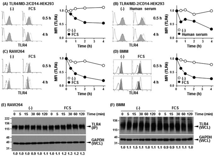
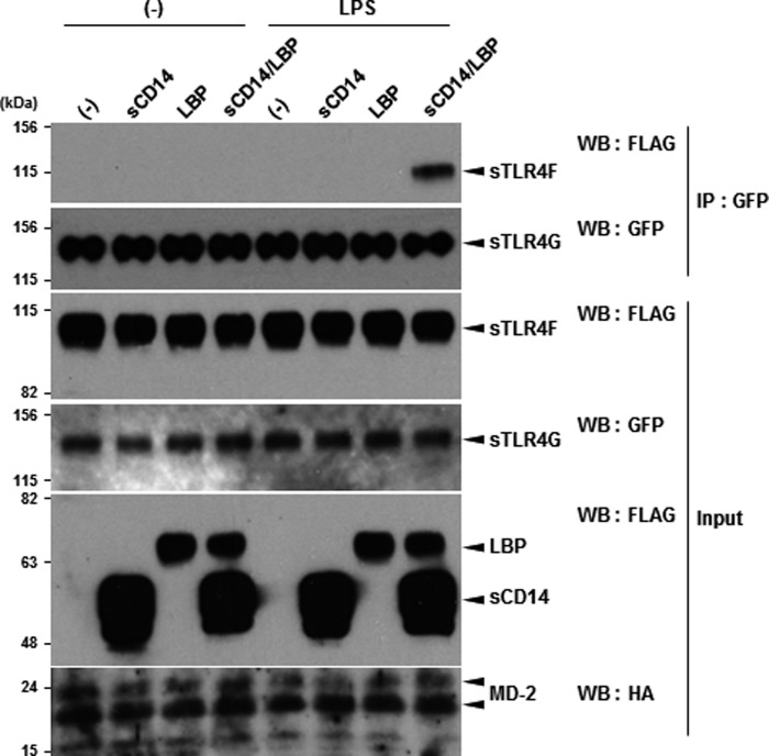
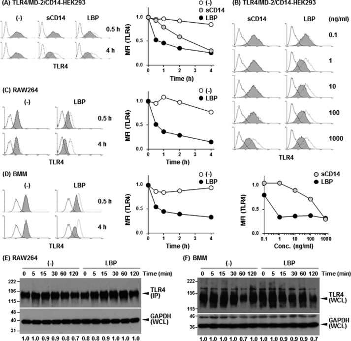
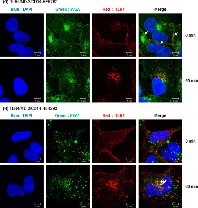
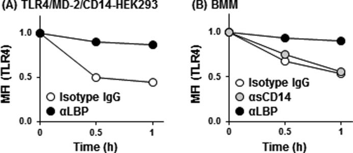
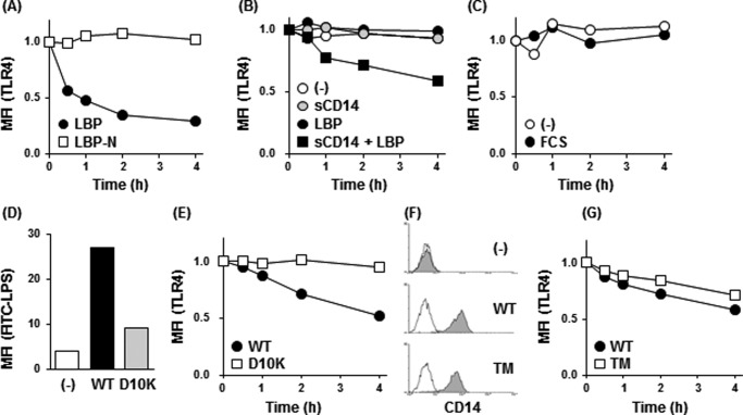
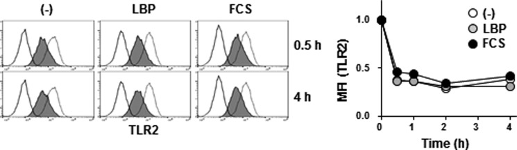
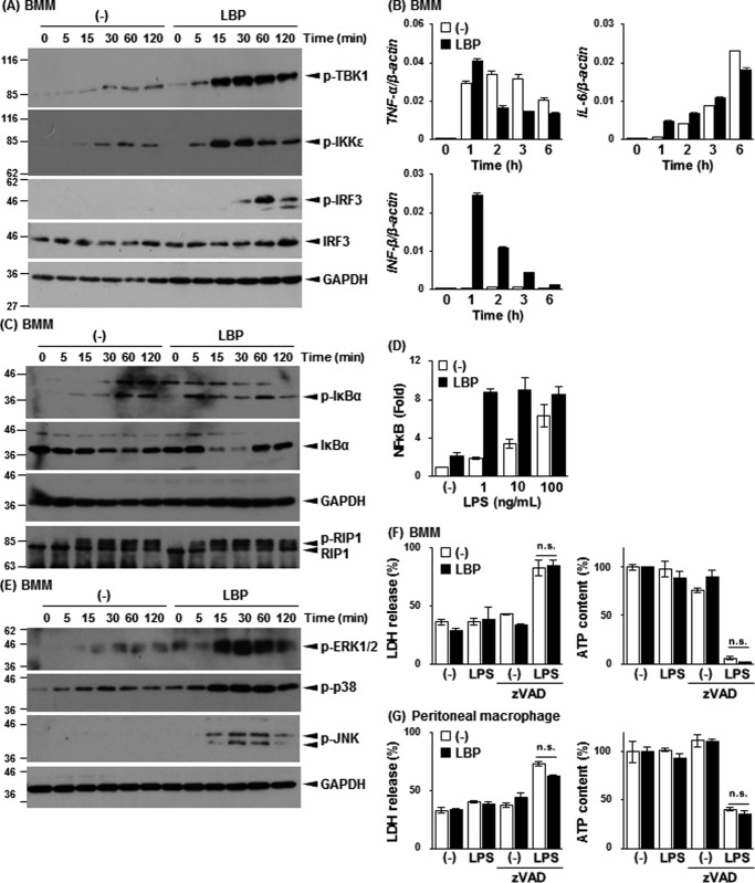
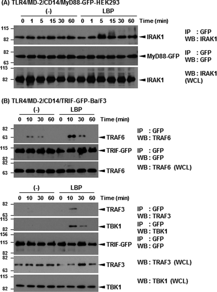
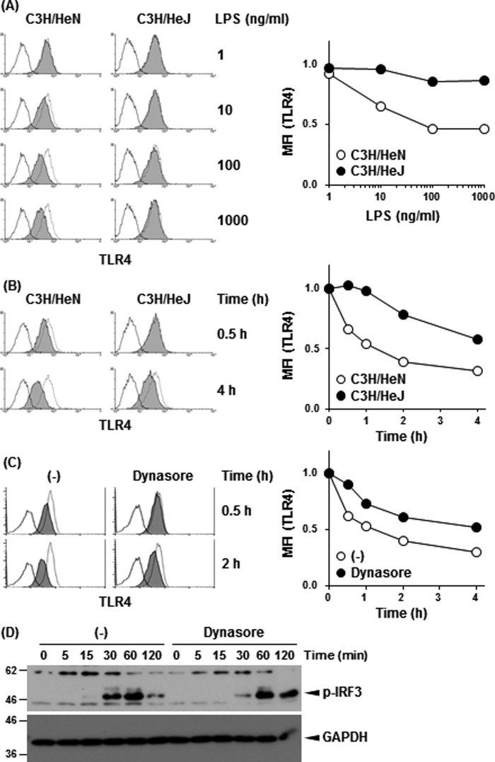
Toll-like receptor 4 (TLR4) is an indispensable immune receptor for lipopolysaccharide (LPS), a major component of the Gram-negative bacterial cell wall. Following LPS stimulation, TLR4 transmits the signal from the cell surface and becomes internalized in an endosome. However, the spatial regulation of TLR4 signaling is not fully understood. Here, we investigated the mechanisms of LPS-induced TLR4 internalization and clarified the roles of the extracellular LPS-binding molecules, LPS-binding protein (LBP), and glycerophosphatidylinositol-anchored protein (CD14). LPS stimulation of CD14-expressing cells induced TLR4 internalization in the presence of serum, and an inhibitory anti-LBP mAb blocked its internalization. Addition of LBP to serum-free cultures restored LPS-induced TLR4 internalization to comparable levels of serum. The secretory form of the CD14 (sCD14) induced internalization but required a much higher concentration than LBP. An inhibitory anti-sCD14 mAb was ineffective for serum-mediated internalization. LBP lacking the domain for LPS transfer to CD14 and a CD14 mutant with reduced LPS binding both attenuated TLR4 internalization. Accordingly, LBP is an essential serum molecule for TLR4 internalization, and its LPS transfer to membrane-anchored CD14 (mCD14) is a prerequisite. LBP induced the LPS-stimulated phosphorylation of TBK1, IKKϵ, and IRF3, leading to IFN-β expression. However, LPS-stimulated late activation of NF-κB or necroptosis were not affected. Collectively, our results indicate that LBP controls LPS-induced TLR4 internalization, which induces TLR adaptor molecule 1 (TRIF)-dependent activation of the TBK1-IKKϵ-IRF3-IFN-β pathway. In summary, we showed that LBP-mediated LPS transfer to mCD14 is required for serum-dependent TLR4 internalization and activation of the TRIF pathway.

Keywords: CD14; LPS-binding protein (LBP); TIR-domain-containing adapter-inducing interferon-B (TRIF); Toll-like receptor 4 (TLR4); cell-surface receptor; endotoxin; innate immunity; lipopolysaccharide (LPS); pathogen-associated molecular pattern (PAMP); pattern recognition receptor (PRR).


Conflict of interest statement

The authors declare that they have no conflicts of interest with the contents of this article

Figures

Figure 1.
Figure 1.
Serum mediates LPS-induced TLR4/MD-2 internalization. A and B, HEK293 cells transfected with human TLR4/MD-2/CD14 were stimulated with LPS (100 ng/ml) with or without 5% FCS (A) or 1% human serum (B). Cells were stained with anti-human TLR4 mAb and PE-conjugated secondary Ab, and analyzed by FACS. The dotted and open histograms represent staining of unstimulated cells with or without primary mAb, respectively. The mean fluorescent intensity (MFI) is presented as the changes relative to unstimulated cells. RAW264 cells (C) and BMMs (D) were stimulated with LPS (100 ng/ml) in the presence or absence of 5% FCS. Cells were stained with biotinylated anti-mouse TLR4 mAb and PE-conjugated Stv and analyzed as in A and B. Data are representative of at least three independent experiments. E, RAW264 cells were stimulated as in C. WCL was subjected to IP with protein G–conjugated anti-TLR4 mAb and then analyzed using Western blotting with anti-TLR4 mAb. As a control for sample preparation, WCL was analyzed using Western blotting with anti-GAPDH mAb. The band intensities of TLR4 normalized to that of GAPDH are represented below the blot images as the changes relative to unstimulated cells in the absence of FBS. F, BMMs were stimulated as in D. WCL was prepared and analyzed using Western blotting with anti-TLR4 and anti-GAPDH, respectively. Data are representative of three independent experiments.
Figure 2.
Figure 2.
LBP mediates LPS-induced dimerization of sTLR4/MD-2 in a CD14-dependent manner. Recombinant sTLR4F/MD-2HA and sTLR4G/MD-2HA were incubated with LPS (5 μg/ml) for 30 min in the presence of FLAG-tagged sCD14, LBP, or both, followed by sTLR4G immunoprecipitation using an anti-GFP Ab-immobilized gel. Precipitated sTLR4F and sTLR4G and the added recombinant proteins (input) were detected by Western blotting using anti-FLAG M2 mAb (sTLR4F, sCD14, and LBP), anti-GFP Ab (sTLR4G), or anti-HA mAb (MD-2), respectively. Data are representative of three independent experiments. WB, Western blotting.
Figure 3.
Figure 3.
Exogenous LBP restored LPS-induced TLR4/MD-2 internalization under the serum-free conditions. A and B, HEK293 cells transfected with human TLR4/MD-2/CD14 were stimulated with LPS (100 ng/ml) in the presence of sCD14 or LBP (1 μg/ml each) (A) or for 4 h in the presence of sCD14 or LBP (B) at the indicated concentrations under the serum-free conditions. TLR4 internalization was analyzed as described in the legend to Fig. 1A. The dotted and open histograms represent staining of unstimulated cells with or without primary mAb, respectively. RAW264 (C) and BMMs (D) were stimulated with LPS (100 ng/ml) in the presence of LBP (100 ng/ml). TLR4 internalization was analyzed as described in the legend to Fig. 1, C and D. Data are representative of at least three independent experiments. RAW264 (E) cells and BMMs (F) were stimulated as in C and D, and total expression of TLR4 was analyzed as described in the legend to Fig. 1, E and F, respectively. Data are representative of three independent experiments. G and H, HEK293 cells transfected with mouse TLR4/MD-2/CD14 were incubated with anti-TLR4 mAb and then stimulated with LPS (1 μg/ml) for 60 min in the presence of LBP (100 ng/ml). G, following stimulation, cells were fixed, stained with CF®488A-conjugated WGA (green), and then permeabilized with 0.1% Triton X-100. Internalized Ab-bound cell-surface TLR4 was reacted with Alexa 546-conjugated F(ab′)2 goat anti-mouse IgG (red). H, cells were fixed, permeabilized, and stained with rabbit anti-EEA1 Ab, followed by Alexa 488-conjugated goat anti-rabbit IgG (green) and Alexa 546-conjugated F(ab′)2 goat anti-mouse IgG (red). Nuclei were counterstained with DAPI (blue). The arrow indicates cell-surface TLR4 on WGA-positive plasma membrane. Arrowhead indicates internalized TLR4 on EEA1-positive endosomes. Original magnification, ×40 objective. Scale bar, 5 μm. Images are representative of two (G) and three (H) independent experiments.
Figure 3.
Figure 3.
Exogenous LBP restored LPS-induced TLR4/MD-2 internalization under the serum-free conditions. A and B, HEK293 cells transfected with human TLR4/MD-2/CD14 were stimulated with LPS (100 ng/ml) in the presence of sCD14 or LBP (1 μg/ml each) (A) or for 4 h in the presence of sCD14 or LBP (B) at the indicated concentrations under the serum-free conditions. TLR4 internalization was analyzed as described in the legend to Fig. 1A. The dotted and open histograms represent staining of unstimulated cells with or without primary mAb, respectively. RAW264 (C) and BMMs (D) were stimulated with LPS (100 ng/ml) in the presence of LBP (100 ng/ml). TLR4 internalization was analyzed as described in the legend to Fig. 1, C and D. Data are representative of at least three independent experiments. RAW264 (E) cells and BMMs (F) were stimulated as in C and D, and total expression of TLR4 was analyzed as described in the legend to Fig. 1, E and F, respectively. Data are representative of three independent experiments. G and H, HEK293 cells transfected with mouse TLR4/MD-2/CD14 were incubated with anti-TLR4 mAb and then stimulated with LPS (1 μg/ml) for 60 min in the presence of LBP (100 ng/ml). G, following stimulation, cells were fixed, stained with CF®488A-conjugated WGA (green), and then permeabilized with 0.1% Triton X-100. Internalized Ab-bound cell-surface TLR4 was reacted with Alexa 546-conjugated F(ab′)2 goat anti-mouse IgG (red). H, cells were fixed, permeabilized, and stained with rabbit anti-EEA1 Ab, followed by Alexa 488-conjugated goat anti-rabbit IgG (green) and Alexa 546-conjugated F(ab′)2 goat anti-mouse IgG (red). Nuclei were counterstained with DAPI (blue). The arrow indicates cell-surface TLR4 on WGA-positive plasma membrane. Arrowhead indicates internalized TLR4 on EEA1-positive endosomes. Original magnification, ×40 objective. Scale bar, 5 μm. Images are representative of two (G) and three (H) independent experiments.
Figure 4.
Figure 4.
Serum LBP is required for LPS-induced TLR4/MD-2 dimerization. A, HEK293 cells transfected with human TLR4/MD-2/CD14 were incubated with anti-LBP or isotype control mAb (1 μg/ml each) for 5 min followed by stimulation with LPS (100 ng/ml) in the presence of 1% human serum. B, BMMs were incubated with anti-LBP (1 μg/ml), anti-sCD14 (10 μg/ml), or isotype control mAb (10 μg/ml) for 5 min and stimulated with LPS (100 ng/ml) in the presence of 1% human serum. TLR4 internalization was analyzed by FACS. Data are representative of three independent experiments.
Figure 5.
Figure 5.
LPS transfer to mCD14 by LBP initiates LPS-induced TLR4/MD-2 internalization. A, HEK293 cells transfected with human TLR4/MD-2/CD14 were stimulated with LPS (100 ng/ml) in the presence of LBP or LBP-N (1 μg/ml each). B and C, HEK293 cells transfected with human TLR4/MD-2 were stimulated with LPS (100 ng/ml) in the presence of sCD14, LBP, or both (1 μg/ml each) (B) or in the presence of 5% FCS (C). TLR4 internalization was analyzed by FACS. D, HEK293 cells expressing TLR4/MD-2 and WT or D10K CD14 were incubated with FITC-conjugated LPS (20 μg/ml) for 30 min in the presence of LBP (1 μg/ml). MFI was also determined by FACS. E, cells in D were stimulated with LPS (100 ng/ml) in the presence of LBP (1 ng/ml). TLR4 internalization was analyzed by FACS. F, HEK293 cells expressing TLR4/MD-2 and WT or TM CD14 were stained with biotinylated anti-CD14 mAb and PE-conjugated Stv, followed by FACS analysis. G, the open histogram represents staining without primary mAb. Cells in F were stimulated with LPS (100 ng/ml) in the presence of LBP (100 ng/ml). TLR4 internalization was analyzed by FACS. Data are representative of three independent experiments.
Figure 6.
Figure 6.
Ligand-induced TLR2 internalization is independent of serum components. BMMs were stimulated with Pam2CSK4 (1 μg/ml) in the presence of LBP (100 ng/ml) or FCS (5%). TLR2 internalization was analyzed by FACS. Cells were stained with anti-mouse TLR2 mAb and allophycocyanin-conjugated secondary Ab, and surface TLR2 levels were analyzed by FACS. The dotted and open histograms represent the staining of unstimulated cells with or without primary mAb, respectively. Data are representative of three independent experiments.
Figure 7.
Figure 7.
LBP mediates LPS-induced TBK1/IKKϵ/IRF3 phosphorylation, but not delayed NF-κB activation and necroptosis. A, C, and E, BMMs were stimulated with LPS (100 ng/ml) in the presence or absence of LBP (100 ng/ml). Following stimulation, cell lysates were prepared and analyzed by Western blotting. Data are representative of at least three independent experiments. B, BMMs were stimulated as in A. The expression of TNFα, IL-6, and IFN-β mRNA was determined using quantitative real-time PCR and normalized to β-actin mRNA. Data are shown as the mean ± S.E. in triplicate measurements. Experiments were conducted three times with similar results. D, mouse TLR4/MD-2/CD14–expressing Ba/F3–transfected cells carrying NF-κB-responsive luciferase reporter genes were stimulated with LPS for 6 h in the presence or absence of LBP (400 ng/ml). Luciferase activity was shown as the mean ± S.D. of the fold-increase against nonstimulated cells in triplicate cultures. F and G, BMMs (F) and peritoneal resident macrophages (G) were stimulated with LPS (100 ng/ml) and Z-VAD (30 μm) for 24 h in the presence or absence of LBP (100 ng/ml). Cytotoxicity was determined by LDH release into the culture medium. Cell viability was determined by intracellular ATP content. Data are representative of three independent experiments. Bars represent the mean ± S.D. Significant differences were determined by the Student's t test; ns, not significant.
Figure 8.
Figure 8.
LBP mediates LPS-induced Myddosome and Triffosome assembly. A and B, HEK293 cells stably expressing TLR4/MD-2/CD14 and MyD88-GFP (A) and Ba/F3 cells stably expressing TLR4/MD-2/CD14 and TRIF-GFP were stimulated with LPS (1 μg/ml) in the presence or absence of LBP (100 ng/ml). Following stimulation, cell lysates were prepared and then subjected to IP with Affi-Gel 10-conjugated anti-GFP mAb. Precipitated samples and WCLs were analyzed using Western blotting with the indicated Abs. Data are representative of three independent experiments.
Figure 9.
Figure 9.
LPS-induced signal from TLR4 enhances but is not required for LBP-mediated internalization. A and B, BMMs from C3H/HeN and C3H/HeJ mice were stimulated with the indicated concentrations of LPS in the presence of LBP (100 ng/ml) for 1 h (A) or with LPS (100 ng/ml) in the presence of LBP (100 ng/ml) for the indicated periods (B). C, BMMs from C57BL/6N mice were treated with Dynasore (80 μm) for 15 min and then stimulated with LPS (100 ng/ml) in the presence of LBP (100 ng/ml). Surface TLR4 levels were analyzed by FACS. The dotted and open histograms represent staining of unstimulated cells with or without primary mAb, respectively. The shaded histograms represent the staining of LPS-stimulated cells with primary mAb. D, BMMs were stimulated as in C for the indicated periods. Cell lysates were prepared and analyzed using WB with indicated Abs. Data are representative of three independent experiments.

Similar articles

Cited by

References

    1. Kawai T., and Akira S. (2011) Toll-like receptors and their crosstalk with other innate receptors in infection and immunity. Immunity 34, 637–650 10.1016/j.immuni.2011.05.006 - DOI - PubMed
    1. Brubaker S. W., Bonham K. S., Zanoni I., and Kagan J. C. (2015) Innate immune pattern recognition: a cell biological perspective. Annu. Rev. Immunol. 33, 257–290 10.1146/annurev-immunol-032414-112240 - DOI - PMC - PubMed
    1. Iwasaki A., and Medzhitov R. (2015) Control of adaptive immunity by the innate immune system. Nat. Immunol. 16, 343–353 10.1038/ni.3123 - DOI - PMC - PubMed
    1. Kawai T., and Akira S. (2010) The role of pattern-recognition receptors in innate immunity: update on Toll-like receptors. Nat. Immunol. 11, 373–384 10.1038/ni.1863 - DOI - PubMed
    1. Lee C. C., Avalos A. M., and Ploegh H. L. (2012) Accessory molecules for Toll-like receptors and their function. Nat. Rev. Immunol. 12, 168–179 10.1038/nri3151 - DOI - PMC - PubMed

Publication types

MeSH terms