

Cellulose Synthase Stoichiometry in Aspen Differs from Arabidopsis and Norway Spruce
- PMID: 29760198
- PMCID: PMC6053019
- DOI: 10.1104/pp.18.00394
Cellulose Synthase Stoichiometry in Aspen Differs from Arabidopsis and Norway Spruce
Abstract
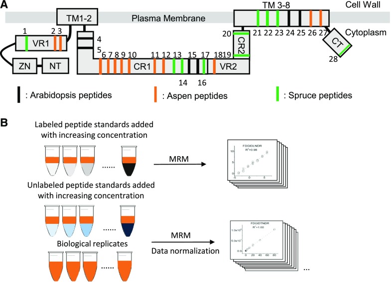
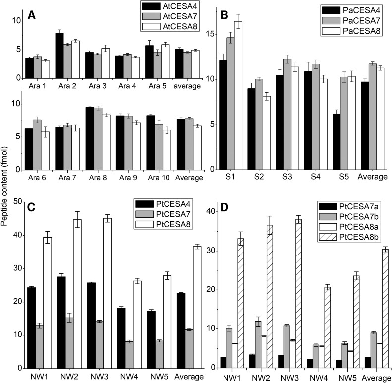
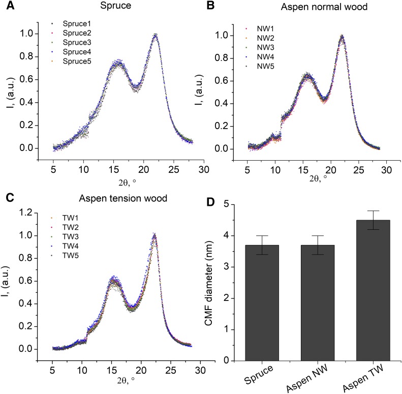
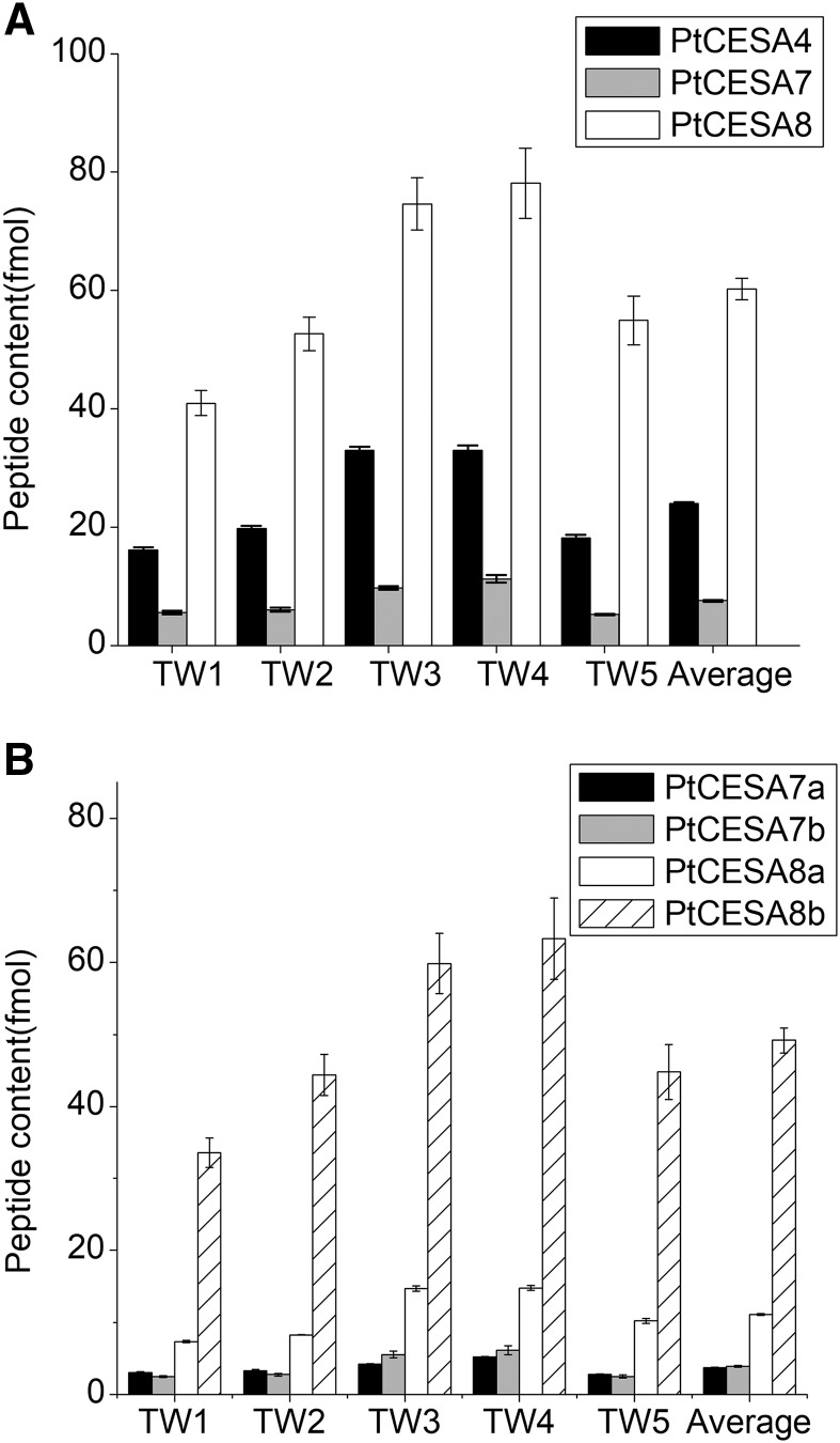
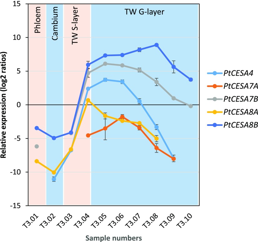
Cellulose is synthesized at the plasma membrane by cellulose synthase complexes (CSCs) containing cellulose synthases (CESAs). Genetic analysis and CESA isoform quantification indicate that cellulose in the secondary cell walls of Arabidopsis (Arabidopsis thaliana) is synthesized by isoforms CESA4, CESA7, and CESA8 in equimolar amounts. Here, we used quantitative proteomics to investigate whether the CSC model based on Arabidopsis secondary cell wall CESA stoichiometry can be applied to the angiosperm tree aspen (Populus tremula) and the gymnosperm tree Norway spruce (Picea abies). In the developing xylem of aspen, the secondary cell wall CESA stoichiometry was 3:2:1 for PtCESA8a/b:PtCESA4:PtCESA7a/b, while in Norway spruce, the stoichiometry was 1:1:1, as observed previously in Arabidopsis. Furthermore, in aspen tension wood, the secondary cell wall CESA stoichiometry changed to 8:3:1 for PtCESA8a/b:PtCESA4:PtCESA7a/b. PtCESA8b represented 73% of the total secondary cell wall CESA pool, and quantitative polymerase chain reaction analysis of CESA transcripts in cryosectioned tension wood revealed increased PtCESA8b expression during the formation of the cellulose-enriched gelatinous layer, while the transcripts of PtCESA4, PtCESA7a/b, and PtCESA8a decreased. A wide-angle x-ray scattering analysis showed that the shift in CESA stoichiometry in tension wood coincided with an increase in crystalline cellulose microfibril diameter, suggesting that the CSC CESA composition influences microfibril properties. The aspen CESA stoichiometry results raise the possibility of alternative CSC models and suggest that homomeric PtCESA8b complexes are responsible for cellulose biosynthesis in the gelatinous layer in tension wood.
© 2018 American Society of Plant Biologists. All rights reserved.
Figures
Comment in
-
Cellulose Synthase Stoichiometry Varies among Species and Tissues.Plant Physiol. 2018 Jul;177(3):873-874. doi: 10.1104/pp.18.00572. Plant Physiol. 2018. PMID: 30006457 Free PMC article. No abstract available.
References
-
- Arioli T, Peng L, Betzner AS, Burn J, Wittke W, Herth W, Camilleri C, Höfte H, Plazinski J, Birch R, et al. (1998) Molecular analysis of cellulose biosynthesis in Arabidopsis. Science 279: 717–720 - PubMed
-
- Brönstrup M. (2004) Absolute quantification strategies in proteomics based on mass spectrometry. Expert Rev Proteomics 1: 503–512 - PubMed
-
- Brown C, Leijon F, Bulone V (2012) Radiometric and spectrophotometric in vitro assays of glycosyltransferases involved in plant cell wall carbohydrate biosynthesis. Nat Protoc 7: 1634–1650 - PubMed
-
- Chiva C, Ortega M, Sabidó E (2014) Influence of the digestion technique, protease, and missed cleavage peptides in protein quantitation. J Proteome Res 13: 3979–3986 - PubMed
Publication types
MeSH terms
Substances
LinkOut - more resources
Full Text Sources
Other Literature Sources
