. 2018 Jul 23;86(8):e00092-18.
doi: 10.1128/IAI.00092-18. Print 2018 Aug.

Specific Binding to Differentially Expressed Human Carcinoembryonic Antigen-Related Cell Adhesion Molecules Determines the Outcome of Neisseria gonorrhoeae Infections along the Female Reproductive Tract

Affiliations

Specific Binding to Differentially Expressed Human Carcinoembryonic Antigen-Related Cell Adhesion Molecules Determines the Outcome of Neisseria gonorrhoeae Infections along the Female Reproductive Tract

Epshita A Islam et al. Infect Immun. .

Abstract

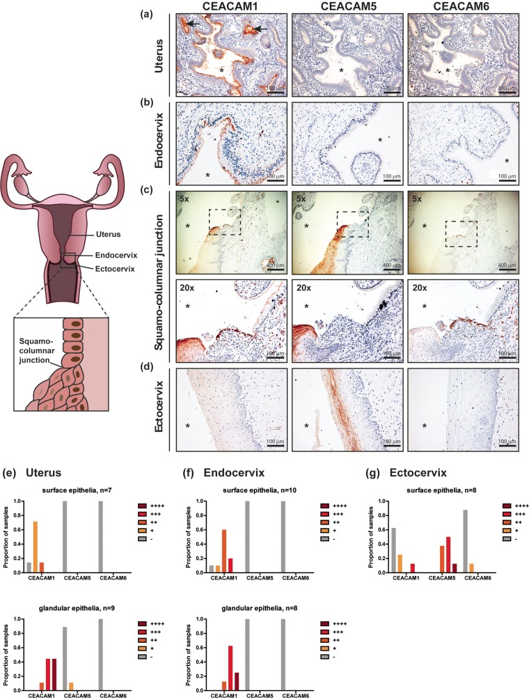
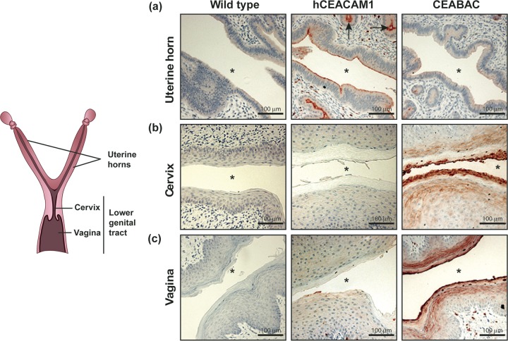
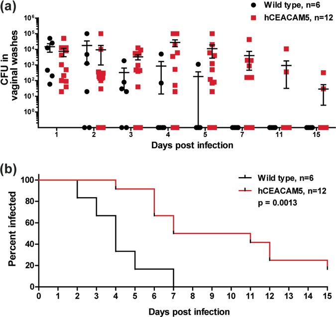
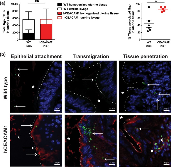
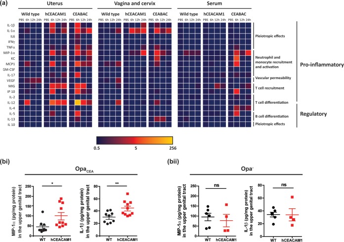
The gonococcal Opa proteins are an antigenically variable family of surface adhesins that bind human carcinoembryonic antigen-related cell adhesion molecule 1 (CEACAM1), CEACAM3, CEACAM5, and/or CEACAM6, cell surface glycoproteins that are differentially expressed on a broad spectrum of human cells and tissues. While they are presumed to be important for infection, the significance of various Opa-CEACAM-mediated cellular interactions in the context of the genital tract has remained unclear. Here, we observed that CEACAM1 and CEACAM5 are differentially expressed on epithelia lining the upper and lower portions of the human female genital tract, respectively. Using transgenic mouse lines expressing human CEACAMs in a manner that reflects this differential pattern, we considered the impact of Opa-CEACAM interactions during uncomplicated lower genital tract infections versus during pelvic inflammatory disease. Our results demonstrate that Opa-CEACAM5 binding on vaginal epithelia facilitates the long-term colonization of the lower genital tract, while Opa protein binding to CEACAM1 on uterine epithelia enhances gonococcal association and penetration into these tissues. While these Opa-dependent interactions with CEACAM-expressing epithelial surfaces promote infection, Opa binding by neutrophil-expressed CEACAMs counterbalances this by facilitating more effective gonococcal clearance. Furthermore, during uterine infections, CEACAM-dependent tissue invasion aggravates disease pathology by increasing the acute inflammatory response. Together, these findings demonstrate that the outcome of infection is determined by both the cell type-specific expression of human CEACAMs and the CEACAM specificity of the Opa variants expressed, which combine to determine the level of gonococcal association with the genital mucosa versus the extent of CEACAM-dependent inflammation and gonococcal clearance by neutrophils.

Keywords: CEACAM1; Neisseria gonorrhoeae; carcinoembryonic antigen (CEA); carcinoembryonic antigen-related cellular adhesion molecules (CEACAM); gonorrhea; inflammation; mucosal infection; pelvic inflammatory disease (PID); sexually transmitted disease (STD).


Figures

FIG 1
FIG 1
Expression of CEACAMs along the human female reproductive tract. (a to d) Hysterectomy samples were stained with mouse monoclonal antibodies raised against human CEACAM1, CEACAM5, or CEACAM6 (red-brown), as indicated. Nuclei were counterstained with hematoxylin (purple). Images were obtained at a ×20 magnification unless otherwise specified. Asterisk, lumen; arrows, uterine glands. (e to g) The CEACAM staining intensity (red-brown) was qualitatively scored to distinguish low (+), moderate (++), strong (+++), and very strong (++++) levels of expression in individual patient samples. The proportion of individuals with the respective levels of each CEACAM is depicted graphically. −, undetectable; n, the number of individual patient samples that were examined for CEACAM expression on surface/glandular epithelia.
FIG 2
FIG 2
Expression of human CEACAMs in the reproductive tract of transgenic mouse lines. Tissues from wild-type mice and two different transgenic mouse lines were stained using a rabbit polyclonal antibody that recognizes human CEACAM1, CEACAM3, CEACAM5, and CEACAM6 (red-brown) but not any mouse CEACAM orthologues. Tissues depicted here were collected from mice at the estrus stage. Nuclei were counterstained with hematoxylin (purple). Asterisk, lumen; arrows, uterine glands. Images were obtained at a ×20 magnification and are representative of those from at least 3 animals.
FIG 3
FIG 3
Contribution of Opa-CEACAM5 interaction on vaginal epithelia during lower genital tract colonization. (a) β-Estradiol-treated wild-type and hCEACAM5 mice were vaginally infected with 107 OpaCEA-expressing N. gonorrhoeae bacteria, and viable gonococci in vaginal lavage samples were enumerated by plating at the indicated time points. The N. gonorrhoeae counts from individual animals and the mean number of N. gonorrhoeae bacteria recovered from all mice (including mice that were still colonized and those that had cleared N. gonorrhoeae) are plotted; error bars represent standard errors. Bacterial counts did not reach statistical significance using a two-way analysis of variance. (b) Graph showing the percentage of mice colonized, as determined on the last day that viable N. gonorrhoeae bacteria could be recovered. The log-rank (Mantel-Cox) test was used to compute the P value, which was statistically significant. Statistical analysis was performed using GraphPad Prism (version 5.04) software.
FIG 4
FIG 4
Contribution of Opa-CEACAM interactions on neutrophils during gonococcal clearance from the lower genital tract. (ai) Immunofluorescence staining of bone marrow-derived neutrophils from wild-type and CEABAC mice infected for 30 min with Opa and OpaCEA-expressing N. gonorrhoeae. N. gonorrhoeae bacteria are stained red, while nuclear DNA is stained with DAPI (blue). Images were taken at a ×63 magnification. (aii) Quantification of wild-type and CEABAC neutrophil-associated OpaCEA and Opa gonococci for 30 to 50 cells per group using microscopy. The bar graph depicts the mean; error bars represent standard deviations. (b) β-Estradiol-treated wild-type and CEABAC mice were vaginally infected with 107 OpaCEA-expressing N. gonorrhoeae bacteria, and viable gonococci in vaginal lavage samples were enumerated by plating at the indicated time points. The graph shows the N. gonorrhoeae counts from individual animals and the mean number of N. gonorrhoeae recovered from all mice (including mice that were still colonized and those that had cleared N. gonorrhoeae); error bars represent standard errors. Bacterial counts did not reach statistical significance using a two-way analysis of variance. (c) Graph showing the percentage of mice colonized, as determined on the last day that viable N. gonorrhoeae bacteria could be recovered. The log-rank (Mantel-Cox) test was used to compute the P value to determine the difference between wild-type and CEABAC mice, which was not statistically significant. (d) Neutrophils from the indicated mice were depleted by systemically administering anti-Gr-1 antibody clone RB6-8C5 prior to infection, and the mean number of N. gonorrhoeae bacteria recovered from homogenized tissues at 2 days postinfection is plotted. Error bars represent standard errors. The log increase in N. gonorrhoeae recovery upon neutrophil depletion in CEABAC mice did not reach statistical significance, as measured by one-way analysis of variance. A one-tailed chi-square test comparing the percentage of culture-positive animals among neutrophil-depleted versus control animals produced a P value of 0.09 and 0.02 for wild-type and CEABAC mice, respectively. Statistical analysis was performed using GraphPad Prism (version 5.04) software.
FIG 5
FIG 5
Opa-hCEACAM1-mediated gonococcal association and invasion of the uterine tissues. (a) The uterine horns of β-estradiol-treated wild-type (WT) and hCEACAM1 mice that were transcervically infected with 107 OpaCEA-expressing N. gonorrhoeae (Ngo) bacteria for 6 h were flushed with PBS++ to remove any nonadherent bacteria. (Left) Uterine lavage and homogenized uterine tissues were cultured to quantify nonadherent and tissue-associated bacteria, respectively. Bars represent means; error bars indicate standard errors. ns, not significant, as determined using two-way analysis of variance. (Right) The percentage of tissue-associated N. gonorrhoeae bacteria was calculated by dividing the number of CFU recovered from homogenized tissues by the total number of CFU in homogenized tissues and uterine lavage samples. Error bars represent standard errors. **, P < 0.05, as determined by a t test. Statistical analysis was performed using GraphPad Prism (version 5.04) software. (b) Bacterial localization in the uterine horn of β-estradiol-treated mice 8 h after transcervical infection with 107 OpaCEA-expressing N. gonorrhoeae bacteria. Sections were immunostained with anti-N. gonorrhoeae (green) and anti-human CEACAM (red) antibodies, while nuclei were counterstained with DAPI (blue). White dashed line, uterine epithelial surface; asterisk, the luminal side. Arrows point at N. gonorrhoeae. Arrowheads point at N. gonorrhoeae associated with CEACAM-positive cells in the uterine stroma. Images are representative of those from 3 animals.
FIG 6
FIG 6
Cytokine response during upper genital tract infections of wild-type and CEACAM-expressing mice. (a) Cytokine levels within homogenized tissues and serum, as determined by Luminex multiplex assays after transcervical infection of β-estradiol-treated mice with 107 OpaCEA-expressing N. gonorrhoeae bacteria for 6 h, 12 h, and 24 h. The heat map depicts the fold change normalized to the value for uninfected PBS-treated controls. Data are for 3 mice of each genotype per time point for infected groups and 2 to 3 mice per genotype for uninfected PBS-treated controls. The heat map was generated in Microsoft Excel software. GM-CSF, granulocyte-macrophage colony-stimulating factor; VEGF, vascular endothelial growth factor. The other abbreviations are defined in the text. (b) MIP-1α and IL-1β levels in the upper genital tract of β-estradiol-treated mice at 24 h postinfection with OpaCEA (bi) and Opa (bii) N. gonorrhoeae. Horizontal bars represent means; error bars represent standard errors. P values were determined by a t test. **, P < 0.01; *, P < 0.05; ns, not significant. For panel bi, data points from two independent experiments were pooled and data are for 9 or 10 mice per genotype. For panel bii, data are for 4 or 5 mice per genotype. Statistical analysis was performed using GraphPad Prism (version 5.04) software.

Similar articles

Cited by

References

    1. Gray-Owen SD, Dehio C, Haude A, Grunert F, Meyer TF. 1997. CD66 carcinoembryonic antigens mediate interactions between Opa-expressing Neisseria gonorrhoeae and human polymorphonuclear phagocytes. EMBO J 16:3435–3445. doi:10.1093/emboj/16.12.3435. - DOI - PMC - PubMed
    1. Johansson L, Rytkonen A, Wan H, Bergman P, Plant L, Agerberth B, Hokfelt T, Jonsson AB. 2005. Human-like immune responses in CD46 transgenic mice. J Immunol 175:433–440. doi:10.4049/jimmunol.175.1.433. - DOI - PubMed
    1. Jennings MP, Jen FE, Roddam LF, Apicella MA, Edwards JL. 2011. Neisseria gonorrhoeae pilin glycan contributes to CR3 activation during challenge of primary cervical epithelial cells. Cell Microbiol 13:885–896. doi:10.1111/j.1462-5822.2011.01586.x. - DOI - PMC - PubMed
    1. Porat N, Apicella MA, Blake MS. 1995. Neisseria gonorrhoeae utilizes and enhances the biosynthesis of the asialoglycoprotein receptor expressed on the surface of the hepatic HepG2 cell line. Infect Immun 63:1498–1506. - PMC - PubMed
    1. Ram S, Cullinane M, Blom AM, Gulati S, McQuillen DP, Monks BG, O'Connell C, Boden R, Elkins C, Pangburn MK, Dahlback B, Rice PA. 2001. Binding of C4b-binding protein to porin: a molecular mechanism of serum resistance of Neisseria gonorrhoeae. J Exp Med 193:281–295. doi:10.1084/jem.193.3.281. - DOI - PMC - PubMed

Publication types

MeSH terms