. 2018 Jun;10(6):599-606.
doi: 10.1038/s41557-018-0039-2. Epub 2018 May 14.

Fragment-derived inhibitors of human N-myristoyltransferase block capsid assembly and replication of the common cold virus

Affiliations

Fragment-derived inhibitors of human N-myristoyltransferase block capsid assembly and replication of the common cold virus

Aurélie Mousnier et al. Nat Chem. 2018 Jun.

Abstract

Rhinoviruses (RVs) are the pathogens most often responsible for the common cold, and are a frequent cause of exacerbations in asthma, chronic obstructive pulmonary disease and cystic fibrosis. Here we report the discovery of IMP-1088, a picomolar dual inhibitor of the human N-myristoyltransferases NMT1 and NMT2, and use it to demonstrate that pharmacological inhibition of host-cell N-myristoylation rapidly and completely prevents rhinoviral replication without inducing cytotoxicity. The identification of cooperative binding between weak-binding fragments led to rapid inhibitor optimization through fragment reconstruction, structure-guided fragment linking and conformational control over linker geometry. We show that inhibition of the co-translational myristoylation of a specific virus-encoded protein (VP0) by IMP-1088 potently blocks a key step in viral capsid assembly, to deliver a low nanomolar antiviral activity against multiple RV strains, poliovirus and foot and-mouth disease virus, and protection of cells against virus-induced killing, highlighting the potential of host myristoylation as a drug target in picornaviral infections.


Conflict of interest statement

Competing Financial Interests Statement

A.B., E.W.T., R.J.L., J.A.H. and J.A.B. are inventors on a patent application describing NMT inhibitors including IMP-1031 and IMP-1088 (Bell, A.S.; Tate, E.W.; Leatherbarrow, R.J.; Hutton, J.A.; Brannigan, J.A., “Compounds and their use as inhibitors of N-myristoyl transferase”, PCT In Appl (2017) WO 2017001812).

Figures

Figure 1
Figure 1. Proposed pathway for generation of infectious rhinovirus particles in an infected host cell.
(a) Rhinovirus polyprotein is synthesized in the host, where it undergoes co-translational N-terminal methionine excision by host methionine aminopeptidase (MetAP) to reveal an N-terminal glycine, followed by N-terminal N-myristoylation by host NMT. (b) Capsid assembly starts with cleavage into VP0, VP3 and VP1 by RV protease, and assembly into protomers; protomers then assemble into pentamers, and then into an icosahedral capsid enclosing the RV RNA genome; finally, VP0 is processed into VP4 and VP2, and mature infectious virions are released.
Figure 2
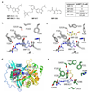
Figure 2. Structure-guided discovery of potent human N-myristoyltransferase inhibitors.
(a) Structures of hit compound IMP-72 and methylated analogue IMP-994, reconstructed fragment IMP-358 and fragment-linked compound IMP-917, and associated inhibitory activity (IC50) on human NMT1 (HsNMT1). (b) Binding mode of IMP-72 in Plasmodium vivax NMT (PvNMT) determined to 1.69 Å resolution, in the presence of NHM (PDB: 5O48). Atoms colored by element: oxygen, red; nitrogen, blue; carbon, ice blue (inhibitor) and grey (protein); fluorine, purple. NHM, protein main chain and solvent molecules have been omitted for clarity, and polar protein-inhibitor interactions are shown by dashed lines. (c) Quaternary complex of PvNMT, NHM, IMP-72 and IMP-358 (PDB: 5O4V) determined to 1.7 Å resolution, with IMP-358 colored in lemon. (d) Binding sites for NHM, IMP-72 and IMP-358, in the context of the overall structure of PvNMT. The protein is a ribbon color-ramped from the N-terminus (blue) to the C-terminus (red). NHM, IMP-72 and IMP-358 are shown in space-filling representation colored by atom type (phosphorus, magenta) and distinguished by carbon atoms shown in grey, ice-blue and lemon, respectively. (e) Binding mode of IMP-917 in human NMT1 (HsNMT1) with Myr-CoA (PDB 5O6H) determined to 1.29 Å resolution. Carbon atoms of the IMP-917 indazole core are colored ice blue, while those of the pendant trimethylpyrazole-containing species are lemon. See Supplementary Fig. 1 for ligand electron density maps.
Figure 3
Figure 3. Potent inhibitors of human N-myristoyltransferases inhibit rhinovirus myristoylation in infected cells.
(a) Structures of IMP-1088 and IMP-1031, and in vitro inhibitory activity (IC50) on human NMT1 (HsNMT1) and NMT2 (HsNMT2). (b) Binding mode of IMP-1088 in the ternary complex with human NMT1 and Myr-CoA (PDB: 5MU6), determined to 1.88 Å resolution, colored as in Fig. 2e. (c) Analysis of protein myristoylation in HeLa cells infected with rhinovirus RV-A16 (MOI 20) in presence of YnMyr, following ligation to TAMRA/Biotin azido capture reagent AzTB; red arrow: band of increased fluorescence intensity upon infection, corresponding to VP0. (d) Quantitative proteomic analysis of inhibition of protein myristoylation in RV-A16-infected HeLa cells (6 hours) by IMP-1088, following ligation to AzRB and enrichment on NeutrAvidin-agarose beads. Two-sample t-test (permutation-based false discovery rate (FDR), 250 permutations, FDR 0.01, S0=0.5) revealed significant changes in label-free quantification (LFQ) between 50 nM IMP-1088 and control (DMSO) for myristoylated proteins. Dashed lines: t-test significance cut off; red: RV-A16 proteins; blue: known co-translationally myristoylated host proteins13; white: other host proteins. Similar analyses were performed for the whole proteome (see Supplementary Fig. 3). (e) Western blot and in-gel fluorescence analysis of inhibition of VP0 myristoylation in HeLa cells infected with rhinovirus RV-A16 (MOI 20, 6 h) following recovery ligation to AzTB. Comparison of input protein, supernatant following pull-down on Streptavidin magnetic beads (Supnt) and eluted proteins (Pull-down) demonstrates enrichment of YnMyr-tagged VP0, and specific inhibition of myristoylation; HSP90: non-myristoylated loading control.
Figure 4
Figure 4. Novel human N-myristoyltransferase inhibitors potently and efficiently block rhinovirus replication without cytotoxicity.
(a) Multicycle rhinovirus replication assay. HeLa cells were infected with rhinovirus RV-A16 (MOI 0.5) for 2 days, and virus replication measured by the induced cytopathic effect (%CPE) for each inhibitor relative to vehicle (DMSO)-treated infected control, and uninfected controls. Error bars: SEM, n=3. (b) Cell viability in presence of NMT inhibitors for uninfected HeLa cells, as percentage of vehicle-treated control. Error bars: SEM, n=3. (c) and (d) Inhibition of single cycle replication in HeLa cells infected with rhinovirus RV-A16 (MOI 20) (c), or the indicated rhinovirus serotypes (d), for 6 h with indicated concentrations of IMP-1088; virus titers determined by endpoint dilution assay. TCID50: 50% Tissue Culture Infective Dose, error bars: SEM, n=4 for (c) and n=3 for (d), ****: p<0.0001 (two-way ANOVA with Sidak's multiple comparisons test on log10(TCID50/ml)). (e) Inhibition of single cycle replication in primary human bronchial epithelial cells (hBECs) infected with rhinovirus RV-A1 (MOI 5) for 7 h by IMP-1088; virus titers determined as in (c)/(d). Error bars: SEM, n=4. (f) Inhibitory effect of IMP-1088 added at different times post-infection, in a single cycle replication assay. HeLa cells were infected with rhinovirus RV-A16 (MOI 20) for 6 h and IMP-1088 (500 nM) added at the indicated times (+/- hours) relative to the time point of virus adsorption. Error bars: SEM, n=3, **: p=0.0013, ****: p<0.0001 (one-way ANOVA with Dunnett's multiple comparisons test on log10(TCID50/ml) against the DMSO control).
Figure 5
Figure 5. IMP-1088 inhibits production of infectious rhinovirus particles by blocking virus assembly.
(a-d) Effect of IMP-1088 on rhinovirus replication kinetics in HeLa cells infected with rhinovirus RV-A16 (MOI 20) for 6 h in presence of IMP-1088 (500 nM) or DMSO (vehicle). At the indicated time points cell, lysates were analyzed for virus titer by endpoint titration (a), viral RNA by quantitative PCR (b) or viral protein levels by Western blot with an antibody directed against rhinovirus non-structural protein 2C ((c) and (d)). (c) Representative images of one of three independent repeats. (d) Quantification of signal corresponding to 2C protein, relative to Lamin B1 loading control, expressed as a percentage of 6 h DMSO control. Error bars: SEM, n=3. (e) Effect of IMP-1088 on rhinovirus capsid assembly. HeLa cells were infected with rhinovirus RV-A16 for 6 h in presence of IMP-1088 (500 nM) or DMSO (vehicle). Cell lysates were sedimented on sucrose density gradients and the fractions analyzed by Western blot with antibodies raised against whole rhinovirus RV-A16 capsids. (f) Densitometric analysis of (e), with indication of fractions in which controls sedimented (intact virion, empty capsid or monomer/pentamer).

References

    1. Ritchie AI, et al. Pathogenesis of Viral Infection in Exacerbations of Airway Disease. Ann Am Thorac Soc. 2015;12(Suppl 2):S115–132. - PubMed
    1. Kieninger E, et al. High rhinovirus burden in lower airways of children with cystic fibrosis. Chest. 2013;143:782–790. - PubMed
    1. Flight WG, et al. Incidence and clinical impact of respiratory viruses in adults with cystic fibrosis. Thorax. 2014;69:247–253. - PubMed
    1. Thibaut HJ, De Palma AM, Neyts J. Combating enterovirus replication: state-of-the-art on antiviral research. Biochem Pharmacol. 2012;83:185–192. - PubMed
    1. Jiang P, Liu Y, Ma HC, Paul AV, Wimmer E. Picornavirus morphogenesis. Microbiol Mol Biol Rev. 2014;78:418–437. - PMC - PubMed

Publication types