. 2018 May 14;50(5):1-15.
doi: 10.1038/s12276-018-0091-4.

CD82 hypomethylation is essential for tuberculosis pathogenesis via regulation of RUNX1-Rab5/22

Affiliations

CD82 hypomethylation is essential for tuberculosis pathogenesis via regulation of RUNX1-Rab5/22

Hyun-Jung Koh et al. Exp Mol Med. .

Abstract

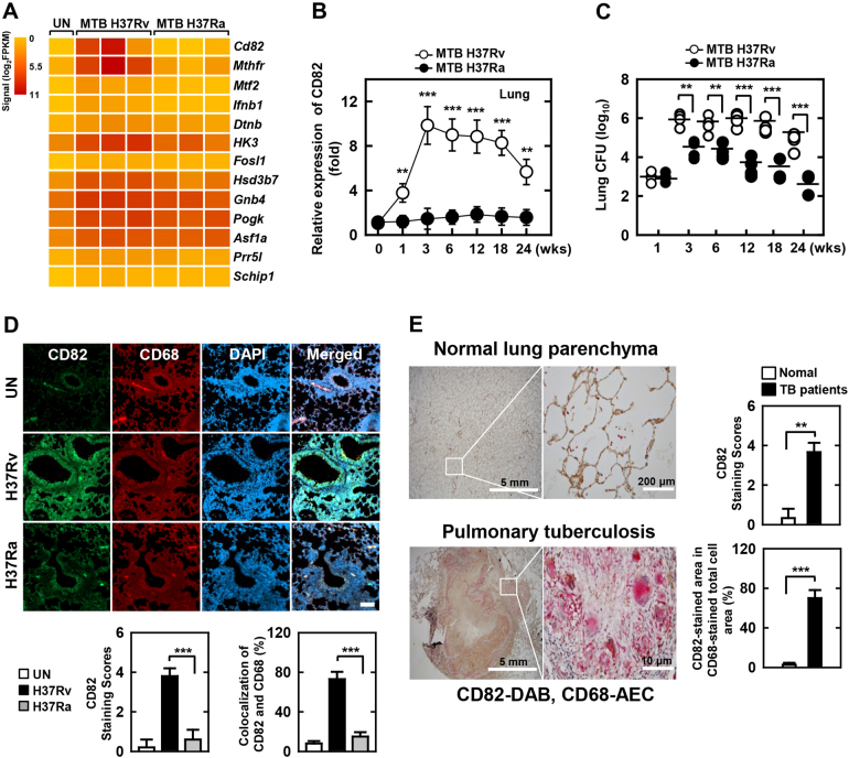
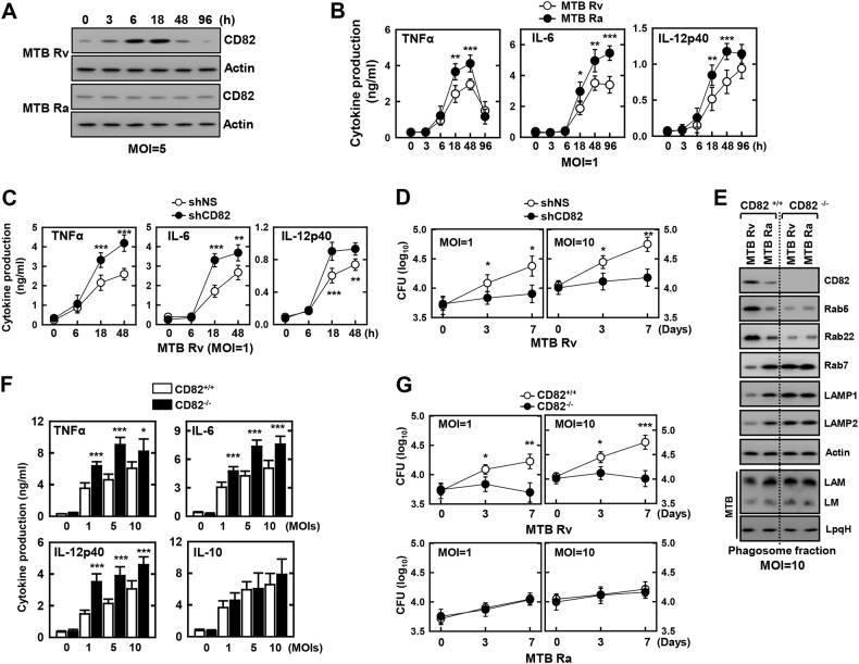
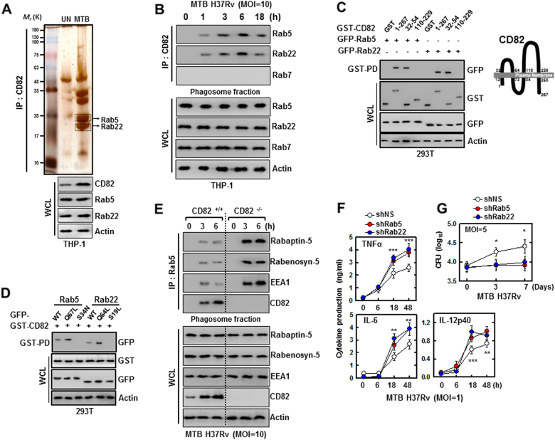
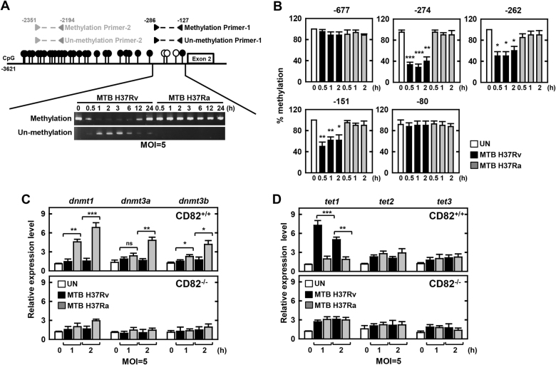
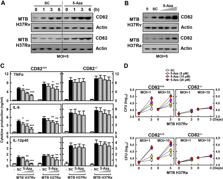
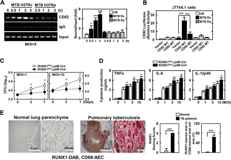
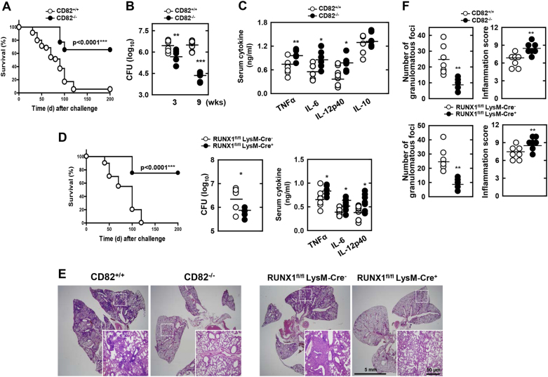
The tumor suppressor gene CD82/KAI1 is a member of the tetraspanin superfamily and organizes various membrane-based processes. Mycobacterium tuberculosis (MTB) persists in host macrophages by interfering with phagolysosome biogenesis and inflammatory responses, but the role of CD82 in controlling the intracellular survival of pathogenic mycobacteria within macrophages remains poorly understood. In this study, we demonstrated that the virulent MTB strain H37Rv (MTB Rv) induced CD82 promoter hypomethylation, resulting in CD82 expression. Targeting of the runt-related transcription factor 1 (RUNX1) by CD82 is essential for phagosome arrest via interacting with Rab5/22. This arrest is required for the intracellular growth of MTB in vitro and in vivo, but not for that of MTB H37Ra (MTB Ra) in macrophages. In addition, knockdown or knockout of CD82 or RUNX1 increased antibacterial host defense via phagolysosome biogenesis, inflammatory cytokine production, and subsequent antimicrobial activity both in vitro and in vivo. Notably, the levels of CD82 and RUNX1 in granulomas were elevated in tuberculosis (TB) patients, indicating that CD82 and RUNX1 have clinical significance in human TB. Our findings identify a previously unrecognized role of CD82 hypomethylation in the regulation of phagosome maturation, enhanced intracellular survival, and the innate host immune response to MTB. Thus, the CD82-RUNX1-Rab5/22 axis may be a previously unrecognized virulence mechanism of MTB pathogenesis.


Conflict of interest statement

The authors declare that they have no conflict of interest.

Figures

Fig. 1
Fig. 1. Identification of MTB Rv-specific CD82 in mice and TB patients.
a Heat map of the top 13 upregulated genes in lungs from MTB Rv- or MTB Ra-infected mice. Each row shows the relative expression level of a single gene, and each column shows the expression level of a single sample. b Real-time qPCR analysis of CD82 expression or c bacterial loads in lungs from MTB Rv- or MTB Ra-intranasal infected mice for the indicated times (as in Fig. S1A). d Representative immunofluorescence images for expression of CD82 and CD68 (a macrophage marker) in lungs from MTB Rv- or MTB Ra-infected mice. Scale bar, 100 μM. The bottom panel shows the quantitative data of staining intensity of CD82 (left) and the colocalization index (%) between CD82 and CD68 (right). e Immunohistochemical analysis to examine CD82-DAB (3,3’-diaminobenzidine) and CD68-AEC (3-amino-9-ethylcarbazole) expression in healthy controls and patients with pulmonary TB. Representative images from five independent healthy controls and patients are shown. Insets, enlargement of outlined areas. Biological replicates (n = 3) for each condition were performed (ad). Significant differences (**P< 0.01; ***P< 0.001) compared with MTB Ra (Student’s t-test with Bonferroni adjustment)
Fig. 2
Fig. 2. The effects of CD82 on cytokine production and bacterial survival in macrophages.
BMDMs were infected with MTB Rv or MTB Ra for the indicated times, followed by a immunoblotting (IB) with αCD82 and αActin, or b culture supernatants were harvested and analyzed for cytokine ELISA for TNFα, IL-6, and IL-12p40. BMDMs were transduced with lentivirus-shRNA-NS or lentivirus-shRNA-CD82 for 2 days (c, d) or BMDMs from CD82+/+ and CD82−/− (f, g) were with MTB Rv or MTB Ra for the indicated times, followed by intracellular survival of MTB assessed by CFU assay (c, f), or culture supernatants were harvested at 18 h and analyzed for cytokine ELISA for TNFα, IL-6, and IL-12p40 (d, g). e BMDMs from CD82+/+ and CD82−/− were infected with MTB Rv or MTB Ra for 6 h. Mycobacteria-containing phagosome fractions were subsequently purified by sucrose-step-gradient ultra-centrifugation, followed by IB to detect αCD82, αRab5, αRab22, αRab7, αLAMP1, αLAMP2, and αActin. The polyclonal MTB Ab and LpqH Ab detect the lipoglycans (LAM and LM) and lipoproteins (LpqH), respectively. The data are representative of four independent experiments with similar results (a, e). Data shown are the means ± SD of six experiments (bd, f, g). Significant differences (*P< 0.05; **P< 0.01; ***P< 0.001) compared with shRNA-NS or CD82+/+ (Student’s t-test with Bonferroni adjustment). CFU colony-forming units
Fig. 3
Fig. 3. CD82 interaction with Rab5 and Rab22 leads to bock maturation.
a Identification of Rab5 and Rab22 by mass spectrometry analysis in THP-1 cells were infected with MTB Rv or MTB Ra (MOI = 5) for 3 h. Whole cell lysates (WCLs) were used for IB with αCD82, αRab5, αRab22, and αActin. b, e THP-1 cells (b) and BMDMs (e) from CD82+/+ and CD82−/− were infected with MTB Rv for the indicated times. Mycobacteria-containing phagosome fractions were subsequently purified by sucrose-step-gradient-ultra-centrifugation, followed by immunoprecipitation (IP) with αCD82 (b) or αRab5 (e) and IB with αRab5, αRab22, αRab7 (b) or αRabaptin-5, αRabenosyn-5, αEEA1, αCD82 (e), and αActin. c Binding mapping. (Left) At 48 h post transfection with mammalian GST or GST-82 and truncated mutant constructs together with GFP-Rab5 or GFP-Rab22, 293T cells were used for GST pulldown, followed by IB with αGFP. WCLs were used for IB with αGST, αGFP or αActin. (Right) Schematic diagrams of the structure of CD82. d 293T cells were co-transfected with GST-CD82 with GFP-Rab5 or GFP-Rab22 and truncated mutant, and subjected to GST pulldown, followed by IB with αGFP. WCLs were used for IB with αGST, αGFP or αActin. f BMDMs were transduced with lentivirus-shRNA-NS or lentivirus-shRNA-Rab5 or Rab22 for 2 days and infected with MTB Rv for the indicated times. Culture supernatants were harvested and analyzed for cytokine ELISA for TNFα, IL-6, and IL-12p40 (f) or intracellular survival of MTB was assessed by CFU assay (g). The data are representative of four independent experiments with similar results (ae). Data shown are the means ± SD of six experiments (f, g). Significant differences (*P< 0.05; **P< 0.01; ***P< 0.001) compared with shRNA-NS (Student’s t-test with ANOVA for multiple comparisons). CFU colony-forming units
Fig. 4
Fig. 4. Methylation analysis of CD82 promoter region during MTB infection.
a Schematic illustration of the CD82 promoter region. The CpG island map is indicated in the top panel, and the sequence amplified for methylation analysis is shown in the bottom panel. BMDMs were infected with MTB Rv or MTB Ra for the indicated times, followed by PCR with methylation or non-methylation primers. b Methylation status of CpG sites was measured by direct sodium bisulfate DNA sequencing as in the Supplementary Methods. Methylation status of −677, −274, −262, −151, or −80 regions was analyzed. At least 10 clones were sequenced for each group. c, d BMDMs from CD82+/+ and CD82−/− were infected with MTB Rv or MTB Ra for the indicated times. Real-time qPCR analysis of dnmt or tet expression. The data are representative of five independent experiments with similar results (a). Data shown are the means ± SD of five experiments (bd). Significant differences (*P< 0.05; **P< 0.01; ***P< 0.001) compared with CD82+/+ (Student’s t-test with Bonferroni adjustment)
Fig. 5
Fig. 5. DNMT inhibition induces CD82 protein expression and anti-mycobacterial responses in a CD82-dependent manner.
BMDMs were pretreated with 5-Aza (a for 10 μM and bd for 5, 10, 20 μM) for 1 h and infected with MTB Rv or MTB Ra for the indicated times, followed by IB with αCD82 and αActin (a, b), culture supernatant harvesting at 18 h and analysis for cytokine ELISA for TNFα, IL-6, and IL-12p40 (c), or intracellular survival of MTB assessment by CFU assay (d). The data are representative of four independent experiments with similar results (a, b). Data shown are the means ± SD of five experiments (c, d). Significant differences (*P< 0.05; **P< 0.01; ***P< 0.001) compared with solvent control (Student’s t-test with Bonferroni adjustment). SC solvent control 0.1% DMSO, CFU colony-forming units
Fig. 6
Fig. 6. CD82 associates with RUNX1.
a ChIP analyses of RUNX1. (Left) BMDMs were infected with MTB Rv for the indicated times, and subjected to semi-PCR analysis using primers specific for CD82. The data are representative of four independent experiments with similar results. (Right) The densitometry results of all four independent RUNX1-CD82 ChIP assays. b J774A.1 cells were transfected with CD82-luciferase reporter constructs (CD82 WT or MT carrying point mutations in the critical RUNX1-binding site) and infected with MTB Rv (MOI = 5) for 6 h. The promoter activities were determined by luciferase assays and normalized to Renilla luciferase enzyme activities. c, d BMDMs from RUNX1fl/fl LysM-Cre+ and RUNX1fl/fl LysM-Cre- mice were infected with MTB Rv for the indicated times, followed by c intracellular survival of MTB assessment by CFU assay or d culture supernatant harvesting at 18 h and analysis by cytokine ELISA for TNFα, IL-6, and IL-12p40. e Immunohistochemical analysis to examine RUNX1-DAB and CD68-AEC expression levels in healthy controls and patients from pulmonary TB. Representative images from 5 independent healthy controls and patients are shown. Insets, enlargement of outlined areas. Data shown are the means ± SD of five experiments (bd). Significant differences (*P< 0.05; **P< 0.01; ***P< 0.001) compared with MTB Ra or RUNX1fl/fl LysM-Cre- (Student’s t-test with Bonferroni adjustment). CFU colony-forming units
Fig. 7
Fig. 7. Modulation of CD82 and RUNX1 affects TB pathogenesis after MTB infection.
a, d Survival of (a) CD82+/+ and CD82−/− mice (n = 35) or (d) RUNX1fl/fl LysM-Cre+ and RUNX1fl/fl LysM-Cre- mice (n = 20) infected with a high intravenous dose (1 × 108 CFUs per mouse) of MTB Rv and monitored for 200 days. Significant differences in comparison to the control mice are indicated (log-rank test). bf CD82+/+ and CD82−/− or RUNX1fl/fl LysM-Cre+ and RUNX1fl/fl LysM-Cre- mice were infected with an intranasal dose (1 × 103 CFUs per mouse) of MTB Rv. b, d Bacterial loads in lungs and c, d serum cytokine levels were determined at 3 weeks; n = 7. e Histopathology scores were obtained from H&E-stained lung sections at 3 weeks, as described in the Methods. Insets, enlargement of outlined areas. f Number of granulomas and inflammation scores observed in seven different lung sections per mouse. Black bars represent the median values. Student’s t-test and Grubbs’ outlier test were used for statistical analysis. The data are representative of two independent experiments with similar results. Significant differences (*P< 0.05; **P< 0.01; ***P< 0.001) compared with CD82+/+ and RUNX1fl/fl LysM-Cre- (Student’s t-test with Bonferroni adjustment). CFU colony-forming units

References

    1. Tonoli H, Barrett JC. CD82 metastasis suppressor gene: a potential target for new therapeutics? Trends Mol. Med. 2005;11:563–570. doi: 10.1016/j.molmed.2005.10.002. - DOI - PubMed
    1. Tsai YC, Weissman AM. Dissecting the diverse functions of the metastasis suppressor CD82/KAI1. FEBS Lett. 2011;585:3166–3173. doi: 10.1016/j.febslet.2011.08.031. - DOI - PMC - PubMed
    1. Odintsova E, et al. Metastasis suppressor tetraspanin CD82/KAI1 regulates ubiquitylation of epidermal growth factor receptor. J. Biol. Chem. 2013;288:26323–26334. doi: 10.1074/jbc.M112.439380. - DOI - PMC - PubMed
    1. Liu WM, Zhang XA. KAI1/CD82, a tumor metastasis suppressor. Cancer Lett. 2006;240:183–194. doi: 10.1016/j.canlet.2005.08.018. - DOI - PubMed
    1. Vyas JM, et al. Tubulation of class II MHC compartments is microtubule dependent and involves multiple endolysosomal membrane proteins in primary dendritic cells. J. Immunol. 2007;178:7199–7210. doi: 10.4049/jimmunol.178.11.7199. - DOI - PMC - PubMed

Publication types

MeSH terms