. 2018 Apr 30:9:846.
doi: 10.3389/fimmu.2018.00846. eCollection 2018.

Podosomes, But Not the Maturation Status, Determine the Protease-Dependent 3D Migration in Human Dendritic Cells

Affiliations

Podosomes, But Not the Maturation Status, Determine the Protease-Dependent 3D Migration in Human Dendritic Cells

Céline Cougoule et al. Front Immunol. .

Abstract

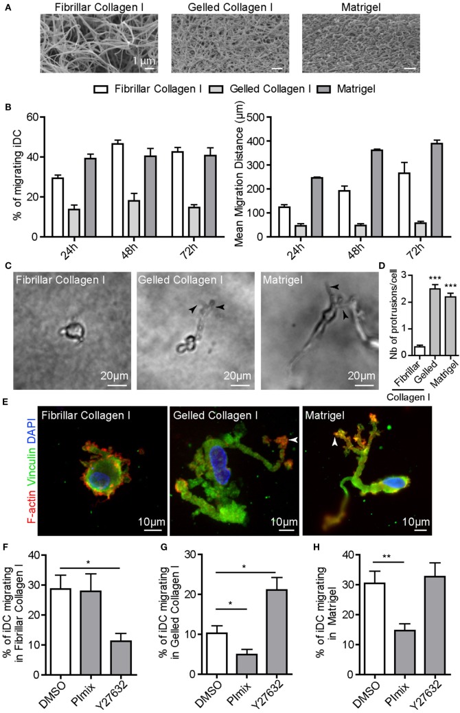
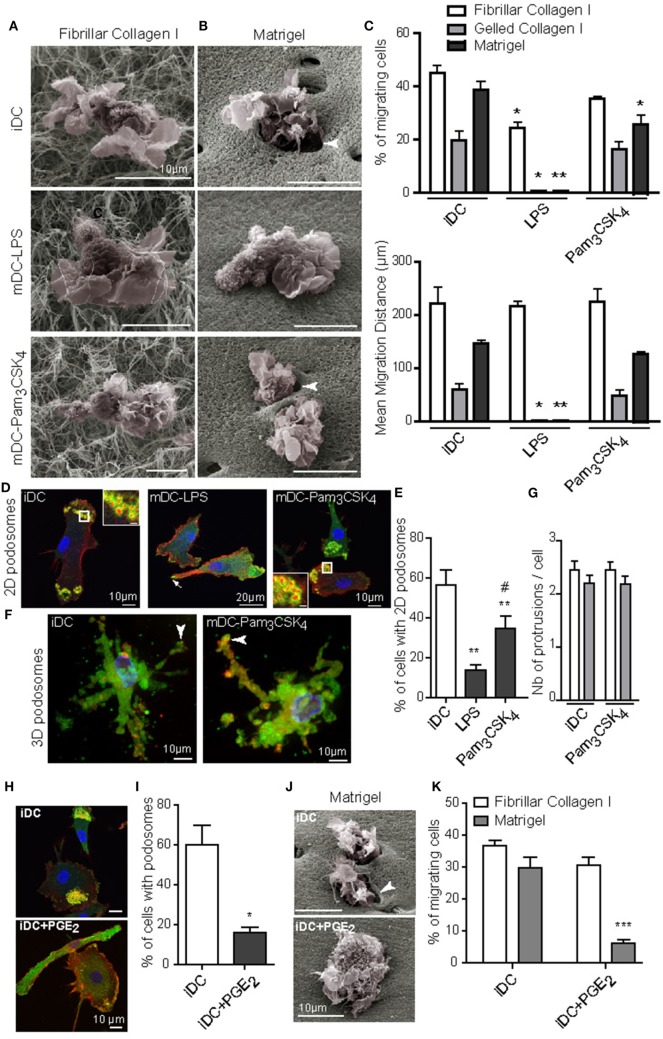
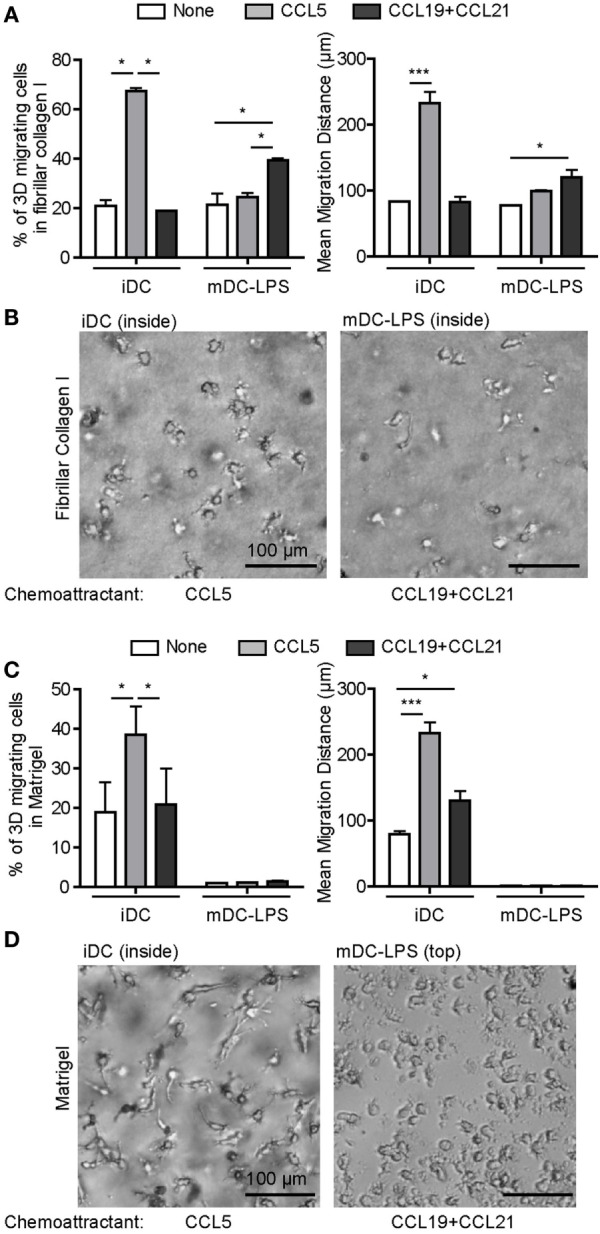
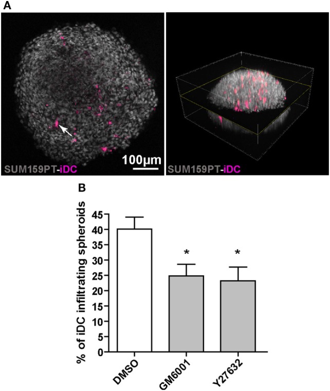
Dendritic cells (DC) are professional Antigen-Presenting Cells scattered throughout antigen-exposed tissues and draining lymph nodes, and survey the body for pathogens. Their ability to migrate through tissues, a 3D environment, is essential for an effective immune response. Upon infection, recognition of Pathogen-Associated Molecular Patterns (PAMP) by Toll-like receptors (TLR) triggers DC maturation. Mature DC (mDC) essentially use the protease-independent, ROCK-dependent amoeboid mode in vivo, or in collagen matrices in vitro. However, the mechanisms of 3D migration used by human immature DC (iDC) are still poorly characterized. Here, we reveal that human monocyte-derived DC are able to use two migration modes in 3D. In porous matrices of fibrillar collagen I, iDC adopted the amoeboid migration mode. In dense matrices of gelled collagen I or Matrigel, iDC used the protease-dependent, ROCK-independent mesenchymal migration mode. Upon TLR4 activation by LPS, mDC-LPS lose the capacity to form podosomes and degrade the matrix along with impaired mesenchymal migration. TLR2 activation by Pam3CSK4 resulted in DC maturation, podosome maintenance, and efficient mesenchymal migration. Under all these conditions, when DC used the mesenchymal mode in dense matrices, they formed 3D podosomes at the tip of cell protrusions. Using PGE2, known to disrupt podosomes in DC, we observed that the cells remained in an immature status and the mesenchymal migration mode was abolished. We also observed that, while CCL5 (attractant of iDC) enhanced both amoeboid and mesenchymal migration of iDC, CCL19 and CCL21 (attractants of mDC) only enhanced mDC-LPS amoeboid migration without triggering mesenchymal migration. Finally, we examined the migration of iDC in tumor cell spheroids, a tissue-like 3D environment. We observed that iDC infiltrated spheroids of tumor cells using both migration modes. Altogether, these results demonstrate that human DC adopt the mesenchymal mode to migrate in 3D dense environments, which relies on their capacity to form podosomes independent of their maturation status, paving the way of further investigations on in vivo DC migration in dense tissues and its regulation during infections.

Keywords: 3D migration; dendritic cells; maturation; podosomes; toll-like receptor.


Figures

Figure 1
Figure 1
Immature DC (iDC) adopt either the amoeboid or the mesenchymal migration mode depending on the matrix architecture. iDC were seeded on top of a thick layer of fibrillar or gelled collagen I, or Matrigel polymerized in culture transwell inserts. (A) Scanning electron microscopy pictures revealing the porous (fibrillar collagen I) versus dense (gelled collagen I and Matrigel) architecture of the matrix. (B) The percentage of migrating cells and the mean migration distance of iDC in fibrillar, gelled collagen I or Matrigel were measured. Results are expressed as mean ± SEM of at least three independent experiments. (C) Bright field images of iDC within matrices were taken using an inverted video microscope and illustrate the round cell morphology in fibrillar collagen I and the elongated and protrusive cell morphology in gelled collagen I and Matrigel. (D) Quantification of the number of membrane protrusions per cells. Results are expressed as mean ± SEM of at least 100 cells counted per conditions. (E) After migration in matrices in Transwell inserts for 24 h, samples were fixed, permeabilized, and stained with anti-vinculin (green), phalloidin Texas-Red (red) and DAPI (blue). iDC form 3D podosomes, F-actin-, vinculin-enriched structures at the tip of membrane protrusions when migrating in dense matrices of gelled collagen I and Matrigel (arrowheads). (F–H), The percentage of iDC migrating in fibrillar collagen I (F), gelled collagen I (G) or Matrigel (H) was measured in control or drug-treated cells (PImix or Y27632). Results are expressed as mean ± SEM of at least three independent experiments.
Figure 2
Figure 2
TLR4 and PGE2, but not TLR2, activation induced podosome dissolution and abolished 3D mesenchymal migration of dendritic cells (DC). Immature DC (iDC) were left untreated or stimulated with LPS (10 ng/mL) or Pam3CSK4 (100 ng/mL) for 16 h. (A,B), Morphology of cells and interaction with the surrounding matrix of fibrillar collagen I (A) or Matrigel (B) were visualized by scanning electron microscopy. Note the presence of holes in the matrix around iDC and mature DC (mDC)-Pam3CSK4 penetrating Matrigel (arrowheads), which were not seen in mDC-LPS. (C) The percentage of migrating cells and the mean migration distance in fibrillar, gelled collagen I or Matrigel, were measured after 24 h. Results are expressed as mean ± SEM of at least three independent experiments. (D) 2D podosomes were stained with an anti-vinculin Ab (green), phalloidin Texas-Red to detect F-actin (red), and DAPI to stain nuclei (blue) (scale bar inset: 2 μm). (E) The percentage of cells forming 2D podosomes was quantified. Results are expressed as mean ± SEM of seven independent experiments. (F) 3D podosomes of iDC and mDC-Pam3CSK4 in gelled collagen I were stained with an anti-vinculin Ab (green), phalloidin Texas-Red to detect F-actin (red), and DAPI to stain nuclei (blue). (G) The number of membrane protrusions per cell was quantified in gelled collagen I (white) and Matrigel (gray). (H–K), iDC were left untreated or treated with PGE2 (5 μM) for 16 h. (H) Podosomes were stained with an anti-vinculin Ab (green), phalloidin Texas-Red to detect F-actin (red), and DAPI to stain nuclei (blue). (I) the percentage of cells forming 2D podosomes was quantified. Results are expressed as mean ± SEM of seven independent experiments. (J) Morphology of cells and interaction with the surrounding matrix of Matrigel were visualized by scanning electron microscopy. Note the presence of holes in the matrix around iDC (arrowhead), which were not seen in iDC + PGE2. (K) The percentage of cells migrating in fibrillar collagen I or Matrigel was measured after 24 h. Results are expressed as mean ± SEM of at least three independent experiments. *P < 0.05; **P < 0.01; ***P < 0.001 compared to iDC condition. #P < 0.05 compared to mDC-LPS condition.
Figure 3
Figure 3
Chemokines regulate three-dimensional (3D) migration of dendritic cells (DC). (A) Immature DC (iDC) and mature DC (mDC)-LPS were seeded on top of a matrix of fibrillar collagen I and the percentage of migrating cells (left) or the mean migration distance (right) were monitored after 6 h, when none (white), CCL5 (gray), or a mixture of CCL19 and CCL21 (black) were added in the lower chamber as chemoattractant. Results are expressed as mean ± SEM of three independent experiments. *P < 0.05; ***P < 0.001 compared to iDC condition. (B) Bright field images of iDC and mDC-LPS within the matrix of fibrillar collagen I were taken using an inverted video microscope and illustrate the round cell morphology in response to CCL5 or a mixture of CCL19 and CCL21 chemokines, respectively. (C) iDC and mDC-LPS were seeded on top of Matrigel and the percentage of 3D migrating cells (left) or the mean migration distance (right) were monitored after 6 h, when none (white), CCL5 (gray), or a mix of CCL19 and CCL21 (black) were added in the lower chamber as chemoattractant. Results are expressed as mean ± SEM of three independent experiments. *P < 0.05; ***P < 0.001. (D) Bright field images of iDC within Matrigel were taken using an inverted video microscope and illustrate the elongated cell morphology in response to CCL5, while mDC-LPS remained on top of the matrix under the influence of CCL19 and CCL21 chemokines.
Figure 4
Figure 4
Immature dendritic cells (iDC) adopt both amoeboid and mesenchymal three-dimensional (3D) migration in tumor spheroids. SUM159PT cell spheroids were coincubated for 3 days with CellTracker-stained DC, with or without drugs. (A) Multiphoton acquisition of DAPI stained spheroids (gray) infiltrated by CellTracker-stained iDC (purple). A spheroid cross-section (left) set in the 3D spheroid reconstitution is shown (right). The arrow indicates a CellTracker-stained iDC located inside the spheroid. (B) Quantification of iDC infiltration into spheroids, with or without inhibitors. Results are expressed as the percentage of iDC inside spheroids (100% corresponds to iDC inside plus iDC at the periphery). Results are expressed as mean ± SEM of five independent experiments. *P < 0.05.

References

    1. Lammermann T, Germain RN. The multiple faces of leukocyte interstitial migration. Semin Immunopathol (2014) 36:227–51.10.1007/s00281-014-0418-8 - DOI - PMC - PubMed
    1. Worbs T, Hammerschmidt SI, Forster R. Dendritic cell migration in health and disease. Nat Rev Immunol (2017) 17:30–48.10.1038/nri.2016.116 - DOI - PubMed
    1. Ng LG, Hsu A, Mandell MA, Roediger B, Hoeller C, Mrass P, et al. Migratory dermal dendritic cells act as rapid sensors of protozoan parasites. PLoS Pathog (2008) 4:e1000222.10.1371/journal.ppat.1000222 - DOI - PMC - PubMed
    1. Farache J, Koren I, Milo I, Gurevich I, Kim KW, Zigmond E, et al. Luminal bacteria recruit CD103+ dendritic cells into the intestinal epithelium to sample bacterial antigens for presentation. Immunity (2013) 38:581–95.10.1016/j.immuni.2013.01.009 - DOI - PMC - PubMed
    1. Faure-Andre G, Vargas P, Yuseff MI, Heuze M, Diaz J, Lankar D, et al. Regulation of dendritic cell migration by CD74, the MHC class II-associated invariant chain. Science (2008) 322:1705–10.10.1126/science.1159894 - DOI - PubMed

Publication types