

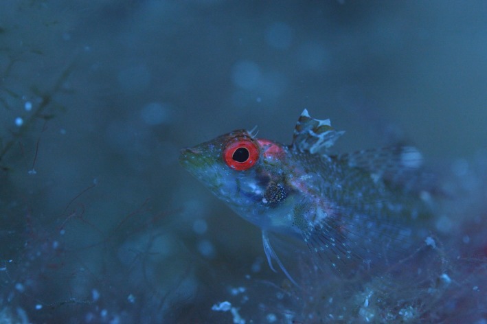
Do the fluorescent red eyes of the marine fish Tripterygion delaisi stand out? In situ and in vivo measurements at two depths
- PMID: 29760908
- PMCID: PMC5938470
- DOI: 10.1002/ece3.4025
Do the fluorescent red eyes of the marine fish Tripterygion delaisi stand out? In situ and in vivo measurements at two depths
Abstract
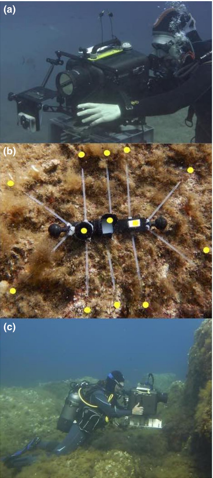
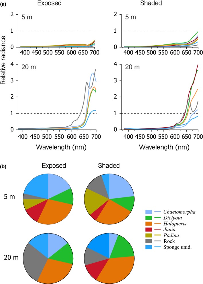
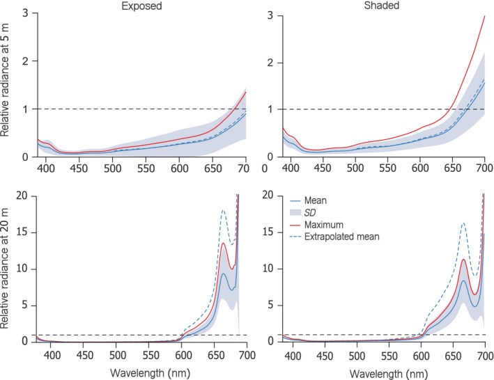
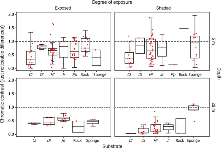
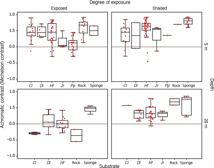
Since the discovery of red fluorescence in fish, much effort has been invested to elucidate its potential functions, one of them being signaling. This implies that the combination of red fluorescence and reflection should generate a visible contrast against the background. Here, we present in vivo iris radiance measurements of Tripterygion delaisi under natural light conditions at 5 and 20 m depth. We also measured substrate radiance of shaded and exposed foraging sites at those depths. To assess the visual contrast of the red iris against these substrates, we used the receptor noise model for chromatic contrasts and Michelson contrast for achromatic calculations. At 20 m depth, T. delaisi iris radiance generated strong achromatic contrasts against substrate radiance, regardless of exposure, and despite substrate fluorescence. Given that downwelling light above 600 nm is negligible at this depth, we can attribute this effect to iris fluorescence. Contrasts were weaker in 5 m. Yet, the pooled radiance caused by red reflection and fluorescence still exceeded substrate radiance for all substrates under shaded conditions and all but Jania rubens and Padina pavonia under exposed conditions. Due to the negative effects of anesthesia on iris fluorescence, these estimates are conservative. We conclude that the requirements to create visual brightness contrasts are fulfilled for a wide range of conditions in the natural environment of T. delaisi.
Keywords: Michelson contrast; coloration; visual communication; visual contrast; visual signal.
Figures
Similar articles
-
Red fluorescence of the triplefin Tripterygion delaisi is increasingly visible against background light with increasing depth.R Soc Open Sci. 2017 Mar 22;4(3):161009. doi: 10.1098/rsos.161009. eCollection 2017 Mar. R Soc Open Sci. 2017. PMID: 28405391 Free PMC article.
-
Fish with red fluorescent eyes forage more efficiently under dim, blue-green light conditions.BMC Ecol. 2017 Apr 20;17(1):18. doi: 10.1186/s12898-017-0127-y. BMC Ecol. 2017. PMID: 28427391 Free PMC article.
-
Controlled iris radiance in a diurnal fish looking at prey.R Soc Open Sci. 2018 Feb 21;5(2):170838. doi: 10.1098/rsos.170838. eCollection 2018 Feb. R Soc Open Sci. 2018. PMID: 29515824 Free PMC article.
-
The consistent difference in red fluorescence in fishes across a 15 m depth gradient is triggered by ambient brightness, not by ambient spectrum.BMC Res Notes. 2016 Feb 17;9:107. doi: 10.1186/s13104-016-1911-z. BMC Res Notes. 2016. PMID: 26887560 Free PMC article.
-
Fluorescent proteins for in vivo imaging, where's the biliverdin?Biochem Soc Trans. 2020 Dec 18;48(6):2657-2667. doi: 10.1042/BST20200444. Biochem Soc Trans. 2020. PMID: 33196077 Review.
Cited by
-
Visual modelling supports the potential for prey detection by means of diurnal active photolocation in a small cryptobenthic fish.Sci Rep. 2019 May 30;9(1):8089. doi: 10.1038/s41598-019-44529-0. Sci Rep. 2019. PMID: 31147614 Free PMC article.
References
-
- Detecting the Detector: A Widespread Animal Sense? Retrieved from http://vixra.org/abs/1411.0226 (Accessed 25.01.2017).
-
- Alieva, N. O. , Konzen, K. A. , Field, S. F. , Meleshkevitch, E. A. , Hunt, M. E. , Beltran‐Ramirez, V. , … Matz, M. V. (2008). Diversity and evolution of coral fluorescent proteins. PLoS ONE, 3(7), e2680 https://doi.org/10.1371/journal.pone.0002680 - DOI - PMC - PubMed
-
- Anthes, N. , Theobald, J. , Gerlach, T. , Meadows, M. G. , & Michiels, N. K. (2016). Diversity and ecological correlates of red fluorescence in marine fishes. Frontiers in Ecology and Evolution, 4, 126.
-
- Anthony, P. D. (1981). Visual contrast thresholds in the cod Gadus Morhua L. Journal of Fish Biology, 19(1), 87–103. https://doi.org/10.1111/j.1095-8649.1981.tb05814.x - DOI
-
- Benjamini, Y. , & Yekutieli, D. (2001). The control of the false discovery rate in multiple testing under dependency. Annals of Statistics, 9, 1165–1188.
LinkOut - more resources
Full Text Sources
Other Literature Sources
