Review
. 2018 Aug;75(16):2963-2980.
doi: 10.1007/s00018-018-2840-x. Epub 2018 May 14.

Imaging of oxygen and hypoxia in cell and tissue samples

Affiliations
Review

Imaging of oxygen and hypoxia in cell and tissue samples

Dmitri B Papkovsky et al. Cell Mol Life Sci. 2018 Aug.

Abstract

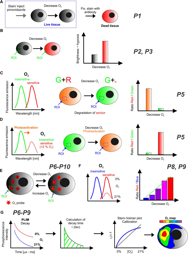
Molecular oxygen (O2) is a key player in cell mitochondrial function, redox balance and oxidative stress, normal tissue function and many common disease states. Various chemical, physical and biological methods have been proposed for measurement, real-time monitoring and imaging of O2 concentration, state of decreased O2 (hypoxia) and related parameters in cells and tissue. Here, we review the established and emerging optical microscopy techniques allowing to visualize O2 levels in cells and tissue samples, mostly under in vitro and ex vivo, but also under in vivo settings. Particular examples include fluorescent hypoxia stains, fluorescent protein reporter systems, phosphorescent probes and nanosensors of different types. These techniques allow high-resolution mapping of O2 gradients in live or post-mortem tissue, in 2D or 3D, qualitatively or quantitatively. They enable control and monitoring of oxygenation conditions and their correlation with other biomarkers of cell and tissue function. Comparison of these techniques and corresponding imaging setups, their analytical capabilities and typical applications are given.

Keywords: FLIM; Fluorescence and phosphorescence-based probes; Fluorescence microscopy; Hypoxia; Live cell and tissue imaging; Oxygen microscopy; PLIM.


Conflict of interest statement

D.B.P. is a former stakeholder of Luxcel Biosciences (now part of Agilent). R.I.D. has no conflicts of interests.

Figures

Fig. 1
Fig. 1
Schemes and signal readouts in optical O2 and hypoxia imaging (see also Table 1). a Redox-sensitive indicators (e.g., pimonidazole) coupled with specific antibody staining of fixed cells/tissue. b Redox-sensitive fluorescent hypoxia stains and fluorescent proteins expressed under HRE promoter. Cells and tissues can be imaged live and fluorescence of different regions of interest (ROI) compared to assess the degree of hypoxia. c Fluorescent proteins with maturation rate sensitive to O2, e.g., UnaG-based [27]. Green fluorescent protein has O2-independent maturation, while the red is not produced under hypoxia. Signal ratio can report on hypoxia. d Red-shifted O2-dependent fluorescence of GFP induced by photoactivation [49]. The ratio between red and green fluorescence reports on hypoxia (reversible changes). eg Quenched phosphorescence and delayed fluorescence-based O2 probes. e O2 probes reversibly change brightness (increases at hypoxia), can be used in semi-quantitative mode shown in b. f Ratiometric O2 probes have two emission bands (e.g., blue and red [, –52]), which allow signal normalization and O2 quantification. g One or two-photon PLIM allows accurate O2 quantification, from measured emission decays and calculated phosphorescence lifetimes (O2 sensitive). Calibration, frequently shown as Stern–Volmer plots, can be linear [53] or non-linear (fitted with the two-site model [54]). PLIM can visualize tissue O2 concentration maps, subcellular gradients, and their dynamics

Similar articles

Cited by

References

    1. James ML, Gambhir SS. A molecular imaging primer: modalities, imaging agents, and applications. Physiol Rev. 2012;92(2):897–965. doi: 10.1152/physrev.00049.2010. - DOI - PubMed
    1. Li Y, Almassalha LM, Chandler JE, Zhou X, Stypula-Cyrus YE, Hujsak KA, Roth EW, Bleher R, Subramanian H, Szleifer I, Dravid VP, Backman V. The effects of chemical fixation on the cellular nanostructure. Exp Cell Res. 2017;358(2):253–259. doi: 10.1016/j.yexcr.2017.06.022. - DOI - PMC - PubMed
    1. Schnell U, Dijk F, Sjollema KA, Giepmans BN. Immunolabeling artifacts and the need for live-cell imaging. Nat Methods. 2012;9(2):152–158. doi: 10.1038/nmeth.1855. - DOI - PubMed
    1. Frigault MM, Lacoste J, Swift JL, Brown CM. Live-cell microscopy–tips and tools. J Cell Sci. 2009;122(6):753–767. doi: 10.1242/jcs.033837. - DOI - PubMed
    1. Stich MIJ, Fischer LH, Wolfbeis OS. Multiple fluorescent chemical sensing and imaging. Chem Soc Rev. 2010;39(8):3102–3114. doi: 10.1039/b909635n. - DOI - PubMed