Review
. 2018 Sep;25(5):353-368.
doi: 10.1097/PAP.0000000000000197.

Recent Advances in the Diagnosis and Pathogenesis of Neurofibromatosis Type 1 (NF1)-associated Peripheral Nervous System Neoplasms

Affiliations
Review

Recent Advances in the Diagnosis and Pathogenesis of Neurofibromatosis Type 1 (NF1)-associated Peripheral Nervous System Neoplasms

Jody F Longo et al. Adv Anat Pathol. 2018 Sep.

Abstract

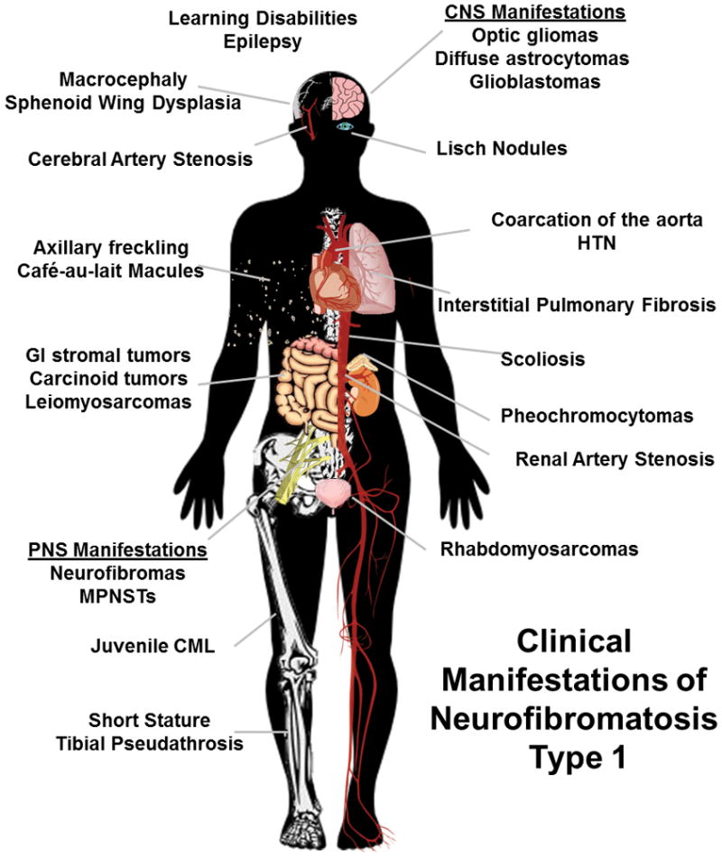
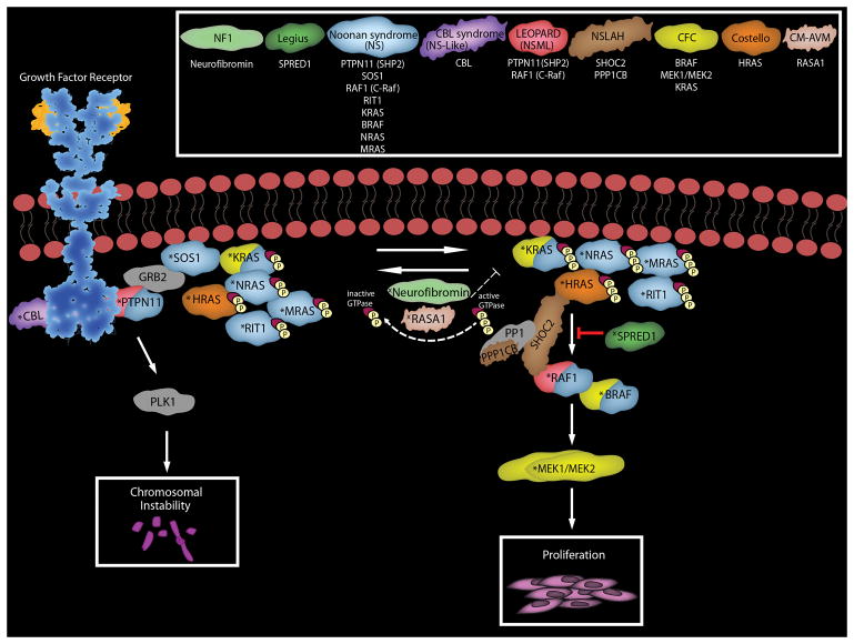
The diagnosis of a neurofibroma or a malignant peripheral nerve sheath tumor (MPNST) often raises the question of whether the patient has the genetic disorder neurofibromatosis type 1 (NF1) as well as how this will impact the patient's outcome, what their risk is for developing additional neoplasms and whether treatment options differ for NF1-associated and sporadic peripheral nerve sheath tumors. Establishing a diagnosis of NF1 is challenging as this disorder has numerous neoplastic and non-neoplastic manifestations which are variably present in individual patients. Further, other genetic diseases affecting the Ras signaling cascade (RASopathies) mimic many of the clinical features of NF1. Here, we review the clinical manifestations of NF1 and compare and contrast them with those of the RASopathies. We also consider current approaches to genetic testing for germline NF1 mutations. We then focus on NF1-associated neurofibromas, considering first the complicated clinical behavior and pathology of these neoplasms and then discussing our current understanding of the genomic abnormalities that drive their pathogenesis, including the mutations encountered in atypical neurofibromas. As several neurofibroma subtypes are capable of undergoing malignant transformation to become MPNSTs, we compare and contrast patient outcomes in sporadic, NF1-associated and radiation-induced MPNSTs, and review the challenging pathology of these lesions. The mutations involved in neurofibroma-MPNST progression, including the recent identification of mutations affecting epigenetic regulators, are then considered. Finally, we explore how our current understanding of neurofibroma and MPNST pathogenesis is informing the design of new therapies for these neoplasms.


Conflict of interest statement

Conflicts of Interest: The authors declare no conflicts of interest.

Figures

FIGURE 1
FIGURE 1
Diagram indicating the neoplastic and non-neoplastic clinical findings potentially encountered in NF1 patients.
FIGURE 2
FIGURE 2
The Ras-associated signaling cascades and the inherited causative genes responsible for the clinical manifestations of RASopathies. RASopathies include neurofibromatosis type 1 (NF1), Legius syndrome, Noonan syndrome (NS), CBL syndrome (Noonan-syndrome like with or without juvenile myelomonocytic leukemia), Noonan syndrome with multiple lentigines (NSML or LEOPARD), Noonan syndrome-like disorder with loose anagen hair (NLSAH), cardio-facio-cutaneous syndrome (CFC), Costello syndrome, and capillary malformation-ateriovenous malformation syndrome (CM-AVM). The most common mutated genes and the resultant proteins associated with each effected disease are color matched as indicated in the key. Note KRAS and BRAF are found mutated in both NS and CFC. Not depicted above are potential causal genes with germline mutations requiring further validation such as SOS2, A2ML1, RASA2, and LZTR1 (Noonan syndrome), RRAS (CBL syndrome) and BRAF (LEOPARD). RASopathy related germline gain-of-function mutations are designated with a “*”, loss-of-function mutations are designated with a “x”. Abbreviations: A2ML1, alpha-2-macroglobulin like 1; BRAF, B-Raf proto-oncogene serine/threonine-protien kinase, CBL, the Cbl proto-oncogene; GRB2, growth factor receptor bound protein 2; LZTR1, leucine zipper like transcription regulator 1; MEK1/MEK2, mitogen-activated protein kinase kinase 1 and 2; neurofibromin, NF1; polo-like-kinase 1 (PLK1); protein phosphatase 1 (PP1), protein phosphatase 1 catalytic subunit beta (PPP1CB); PTPN11, protein tyrosine phosphatase non-receptor typ3 11; RAF1, Raf-1 proto-oncogene; RASA1, Ras p21 protein activator 1; RASA2, Ras p21 protein activator 2; RIT1, Ras-Like without CAAX 1; SHOC2, SHOC2 leucine rich repeat scaffold protein; SOS1, SOS Ras/Rac guanine nucleotide exchange factor 1; SOS2, SOS Ras/Rho guanine nucleotide exchange factor 2; SPRED1, sprout related EVH1 domain containing 1.
FIGURE 3
FIGURE 3
Cellular composition of neurofibromas. A, hematoxylin and eosin stained section of a dermal neurofibroma (40x). All subsequent images are taken from this same tumor at a 63x magnification. B, Immunoreactivity for the transcription factor Sox10 (green, representative cell indicated by an arrow) in the Schwann cell component of this dermal neurofibroma. The section has been counterstained with bisbenzamide to highlight the nuclei of all cells in the tumor. C, Immunoreactivity for the mast cell marker CD117 (also known as c-Kit; red, representative cell indicated by arrow) in the dermal neurofibroma. D, Immunoreactivity for CD34 (red) is apparent in vasculature (arrow) and in small dendritic cells in the tumor. E, Immunoreactivity (red) for the fibroblast nuclear marker TCF4 (transcription factor 4; representative cell indicated by arrow) in the tumor. F, Immunoreactivity for the pan-macrophage marker Iba1 (red, representative cell indicated by arrow) in the tumor. G, Immunoreactivity for CD163 (red), a marker of the M2 (anti-inflammatory) subclass of macrophages, in the tumor (representative cell indicated by arrow). H, Immunoreactivity for CD86 (red), a marker of the M1 (pro-inflammatory) subclass of macrophages, in the tumor (representative cell indicated by arrow).
FIGURE 4
FIGURE 4
Pathology of a MPNST that arose from a plexiform neurofibroma involving the brachial plexus of a 32 year old Caucasian man with NF1. A, section of the MPNST, which is evident as a markedly hypercellular spindle cell neoplasm. The arrow indicates one of the numerous mitotic figures that were detected in this malignancy. B, regions of the original plexiform neurofibroma were readily detected in this resection specimen.

References

    1. Carroll SL, Ratner N. How does the Schwann cell lineage form tumors in NF1? Glia. 2008;56:1590–605. - PMC - PubMed
    1. Carroll SL. Molecular mechanisms promoting the pathogenesis of Schwann cell neoplasms. Acta Neuropathol. 2012;123:321–48. - PMC - PubMed
    1. Shen MH, Harper PS, Upadhyaya M. Molecular genetics of neurofibromatosis type 1 (NF1) J Med Genet. 1996;33:2–17. - PMC - PubMed
    1. Williams VC, Lucas J, Babcock MA, Gutmann DH, Korf B, Maria BL. Neurofibromatosis type 1 revisited. Pediatrics. 2009;123:124–33. - PubMed
    1. Jadayel D, Fain P, Upadhyaya M, Ponder MA, Huson SM, Carey J, Fryer A, Mathew CG, Barker DF, Ponder BA. Paternal origin of new mutations in von Recklinghausen neurofibromatosis. Nature. 1990;343:558–9. - PubMed

MeSH terms

Substances